Lumbar Spine MRI for Back Pain & Sciatica: What to Expect
A lumbar spine MRI is a non-invasive diagnostic scan using powerful magnets and radio waves to create detailed images of the lower back. It helps doctors identify herniated discs, nerve compression, and other causes of chronic pain or sciatica, typically taking 20 to 45 minutes to complete.
If you have been living with persistent lower back pain or the sharp, radiating leg pain known as sciatica, your doctor has likely recommended a lumbar spine MRI. While the prospect of lying in a large machine can feel a bit daunting, understanding the process can significantly reduce any anxiety. At MRI Plus, we believe that transparency is key to a positive patient experience. This guide will walk you through exactly what happens before, during, and after your scan so you can walk into our clinic in London or Leigh on Sea with total confidence.
What exactly happens during a lumbar spine MRI?
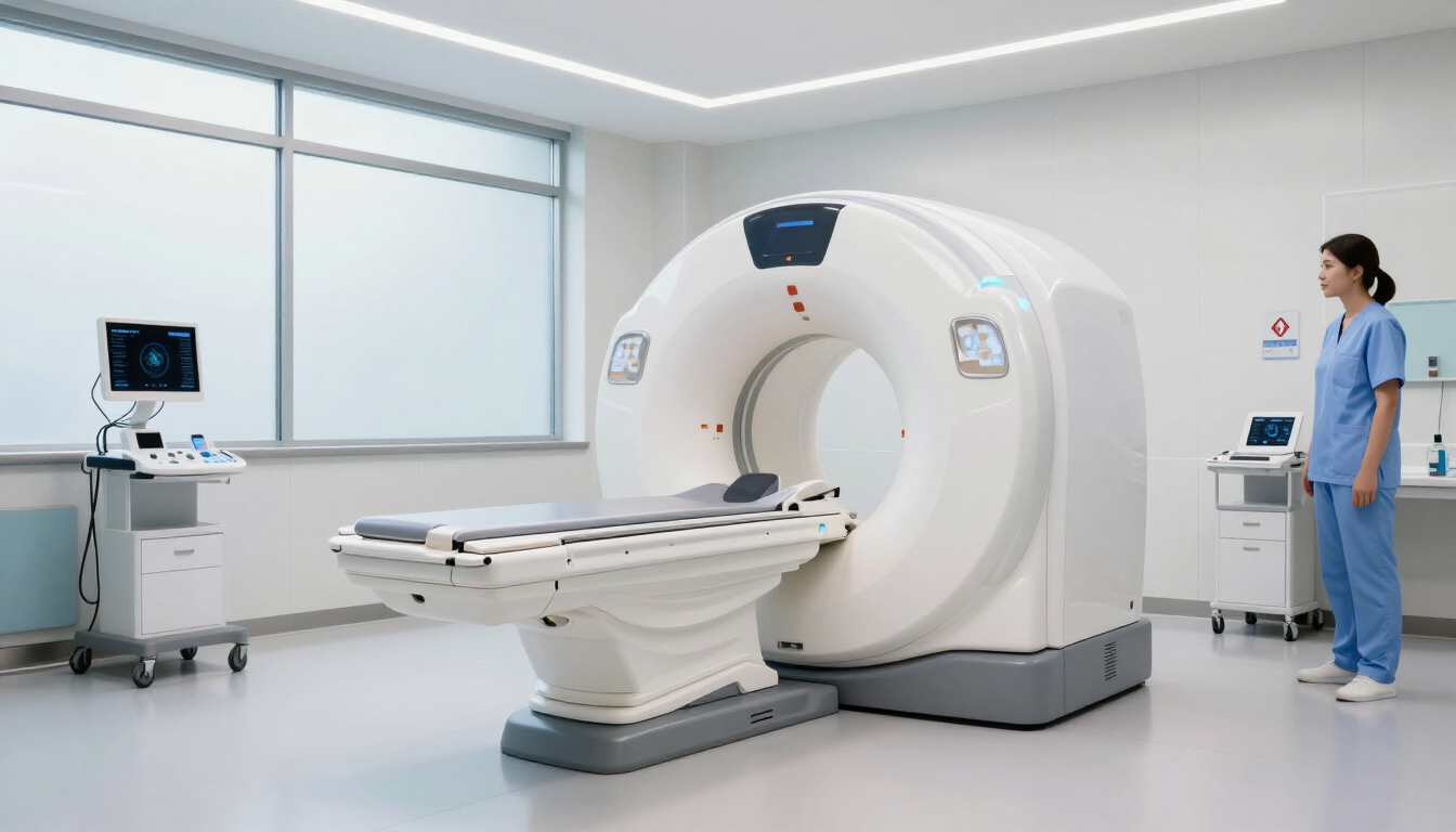
When you arrive for your lumbar spine MRI, the process is streamlined to ensure you spend as little time as possible in the waiting room. After checking in, you will be guided to a private changing area. Because the MRI uses incredibly strong magnets, you cannot have any metal on your body. You may be asked to change into a clinical gown, or you can wear your own comfortable, metal-free clothing (like leggings or joggers without zippers).
Once ready, a radiographer will lead you into the scanning room. You will lie down on a motorised table that slides into the centre of the MRI scanner. For a lumbar scan, you usually go in feet-first, which helps many people feel less enclosed. To get the clearest possible images of your lower vertebrae and discs, a special device called a "coil" might be placed over your abdominal area; this acts as an antenna to capture the radio signals from your body.
During the scan, the most noticeable thing is the noise. The machine makes a series of loud thumping, tapping, and whirring sounds. These are perfectly normal—they are the sounds of the magnetic coils switching on and off to create the image slices. We provide earplugs or headphones with music to help mask the noise. Throughout the procedure, you will be able to speak to the radiographer via an intercom, and you will have a call button in your hand should you need to stop at any point.

Why is a lumbar spine MRI necessary for sciatica?
Sciatica isn't a condition in itself, but rather a symptom of an underlying issue affecting the sciatic nerve. This nerve is the longest in your body, running from your lower back down through your hips and into each leg. When something presses on or irritates this nerve, it causes that characteristic burning or stabbing pain. A lumbar spine MRI is the "gold standard" for diagnosing the cause because it provides far more detail than a standard X-ray.
While an X-ray can show bone issues like fractures or severe arthritis, it cannot see the soft tissues where most sciatica problems reside. The MRI allows clinicians to see:
- Herniated Discs: Where the soft inner material of a spinal disc has leaked out and is pressing on a nerve root.
- Spinal Stenosis: A narrowing of the spaces within your spine, which can put pressure on the nerves.
- Degenerative Disc Disease: Wear and tear of the discs that act as cushions between your vertebrae.
- Spondylolisthesis: When one vertebra slips forward over the one below it.
- Nerve Root Compression: Identifying exactly which nerve is being pinched and where.
How do you prepare for your lower back MRI?
Preparation for a lumbar spine MRI is actually very simple, but there are a few critical safety rules to follow. Because the magnetic field is always active, the most important preparation involves scanning your body for metal. This includes internal medical devices as well as external items.
You should inform the team at MRI Plus if you have any of the following:
- A pacemaker or artificial heart valve
- Cochlear (ear) implants
- Brain aneurysm clips or metallic stents
- Joint replacements or metal plates/screws
- Fragmented metal in your eyes (often from welding or metalwork)
In terms of daily routine, you can generally eat, drink, and take your usual medications as normal. There are no dietary restrictions for a standard lumbar scan unless your doctor has specifically requested a "contrast" MRI. In some cases, a special dye called gadolinium is injected into a vein to highlight certain areas, but this is less common for routine back pain investigations. If you do require contrast, we will provide specific instructions ahead of your appointment.
On the day of the scan, it is best to leave jewelry and watches at home. Even things like hair clips, underwire bras, and some makeup (which can contain trace metallic particles) can interfere with the image quality or cause safety concerns. We suggest arriving 15 minutes early to complete a safety questionnaire and discuss any concerns you might have with our clinical staff.

Key advantages of choosing a private diagnostic provider
When dealing with chronic back pain, time is of the essence. Waiting weeks or even months for a diagnostic scan can lead to worsening symptoms and prolonged physical distress. This is where a private provider like MRI Plus makes a significant difference. Our model is built around speed and accessibility without compromising on clinical excellence.
Choosing a private lumbar spine MRI offers several benefits:
- Rapid Appointment Availability: We aim to offer appointments within days, not weeks, allowing you to get answers faster.
- Prompt Reporting: Our consultant radiologists provide detailed reports quickly, ensuring your GP or specialist can move forward with a treatment plan.
- Convenient Locations: With clinics in London and Leigh on Sea, we provide high-quality care close to home.
- Modern Technology: We use state-of-the-art imaging equipment to ensure the highest resolution for accurate diagnosis.
- Patient Comfort: Our facilities are designed to be calm and welcoming, reducing the stress often associated with hospital environments.
For more information on our latest clinical updates and health tips, you can visit the MRI Plus page.
Can a Lumbar Spine MRI Show Cancer?
Understanding your lower back MRI results
Once the scan is complete, you won't get the results immediately. The "pictures" taken during the session are hundreds of digital cross-sections of your spine. These need to be meticulously reviewed by a Consultant Radiologist—a doctor who specialises in interpreting medical images. They will look at the alignment of your vertebrae, the hydration and height of your discs, and the integrity of the spinal canal.
It is important to remember that MRI findings must always be correlated with your clinical symptoms. Many people have "bulging discs" on an MRI but experience no pain at all. Conversely, a small herniation in a very specific spot can cause debilitating sciatica. Your radiologist will produce a report that describes any abnormalities in detail. This report is then sent to your referring clinician, who will discuss the findings with you and decide on the next steps, which might include physiotherapy, pain management injections, or, in some cases, surgery.
If you are a self-pay patient, we ensure that your results are delivered in a format that is easy for your healthcare provider to use. Our goal is to provide the clinical roadmap you need to regain your mobility and quality of life.
Essential safety protocols for magnetic resonance imaging
Safety is our highest priority at MRI Plus. The magnetic field used in a lumbar spine MRI is thousands of times stronger than a typical kitchen magnet. While this is entirely safe for the human body (there is no ionizing radiation like in a CT scan or X-ray), it means that anything ferromagnetic can become a projectile or heat up.
Our safety protocols include:
- The Screening Questionnaire: A double-check of all implants and medical history.
- Metal Detection: A final sweep before entering the scan room.
- Supervised Access: Only trained clinical staff are permitted within the magnetic zone.
- Constant Monitoring: The radiographer watches and listens to you throughout the entire scan.
By adhering to these strict guidelines, we ensure that every patient has a safe, effective, and stress-free experience. If you suffer from claustrophobia, please let us know in advance. Our modern machines are more spacious than older models, and our staff are experts at helping patients feel relaxed and supported through the process.

Summary and Next Steps
Yes, A lumbar spine MRI can help detect certain types of cancer affecting the lower spine and surrounding tissues.
A lumbar spine MRI is a vital tool for anyone suffering from chronic low back pain or sciatica, providing the clarity needed for an accurate diagnosis. The procedure is safe, painless, and relatively quick, taking about 30-45 minutes. By choosing MRI Plus, you benefit from rapid appointments, expert reporting, and a patient-centric approach that prioritises your comfort.
To move forward with your recovery, consider the following takeaways:
- Confirm your symptoms: Discuss with your doctor if an MRI is the right diagnostic path for your specific pain.
- Check for metal: Ensure you are eligible for an MRI by reviewing any internal medical devices.
- Choose speed: Avoid long waiting lists by opting for a private diagnostic scan.
- Prepare for your visit: Wear comfortable, metal-free clothing on the day of your appointment.
Ready to get answers about your back pain? Contact MRI Plus today to book your lumbar spine MRI and take the first step toward a pain-free life.
References :
Lumbar Spine MRI - I-MED Radiology. (n.d.). https://i-med.com.au/procedures/lumbar-spine-mri
MRI Plus. Whole Spine MRI: How It Works, What It Shows & When It's Used. Available at: https://www.blog.mriplus.co.uk/mri-screening-of-whole-spine
Sklar, S. (2022, December 13). What does a lumbar spine MRI show your doctor? Charlotte Radiology. https://www.charlotteradiology.com/blog/what-does-a-lumbar-spine-mri-show-your-doctor/