Brain MRI: How It Works & What to Expect
A brain MRI is a medical imaging test that uses a powerful magnetic field and radio waves to create detailed 3D images of the brain. It is a non-invasive test. It is mainly used for diagnosis of neurological disorders, detection of structural abnormalities and monitoring treatments without use of harmful ionising radiation .
What is a brain MRI scan used for?
A brain MRI is one of the most versatile and powerful diagnostic tools available in modern medicine. Because the brain is an incredibly complex organ encased in a hard skull, traditional X-rays often struggle to provide a clear view of its soft tissues. This is where Magnetic Resonance Imaging (MRI) shines. By mapping the water molecules in your brain cells, the scanner can produce high-contrast images that reveal even the smallest irregularities.
Clinicians use brain imaging for a wide variety of reasons, ranging from investigating chronic symptoms to ruling out life-threatening emergencies. If you have been experiencing persistent headaches, dizziness, or changes in your vision, your doctor may request a scan to get a better look at what’s happening beneath the surface.
These are the common reasons for a brain scan include:
- Diagnosing migraines and chronic or severe headaches.
- Examination for evidence of a previous stroke or transient ischaemic attack (TIA).
- Detecting and monitoring tumours or cysts in the skull.
- Assessing inflammatory diseases such as Multiple Sclerosis (MS).
- Vascular problems such as aneurysms or malformations (screening).
Understanding the brain imaging process
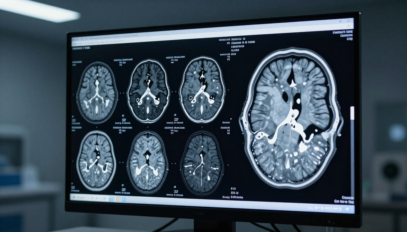
When you think of a medical scan, you might imagine a quick "snapshot." However, a brain MRI is more like a detailed digital dissection. The machine creates "slices" of the brain from various angles—horizontal, vertical, and side-on. These slices are then reconstructed by a computer to create a comprehensive 3D map.
The technology relies on the magnetic properties of hydrogen atoms in your body. When you enter the strong magnetic field of the MRI scanner, these atoms align in a specific direction. Radio waves then temporarily disrupt this alignment. As the atoms return to their original position, they emit signals that the scanner picks up. Different tissues (like white matter, grey matter, and cerebrospinal fluid) emit different signals, allowing the computer to tell them apart with incredible precision.
When is a brain MRI necessary?
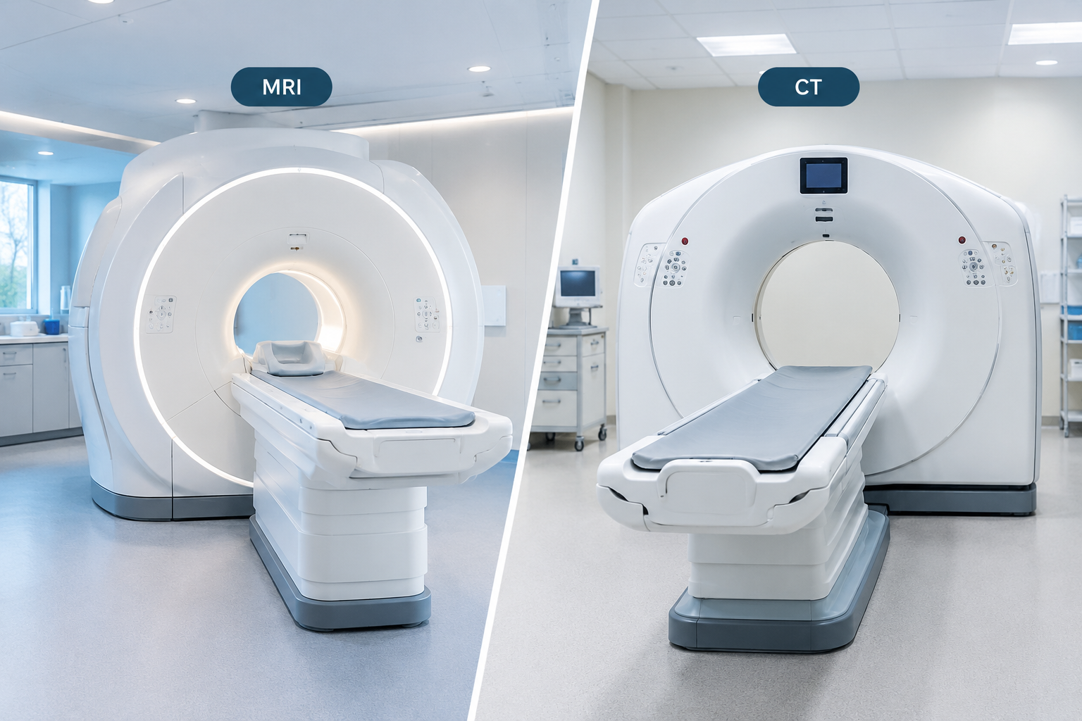
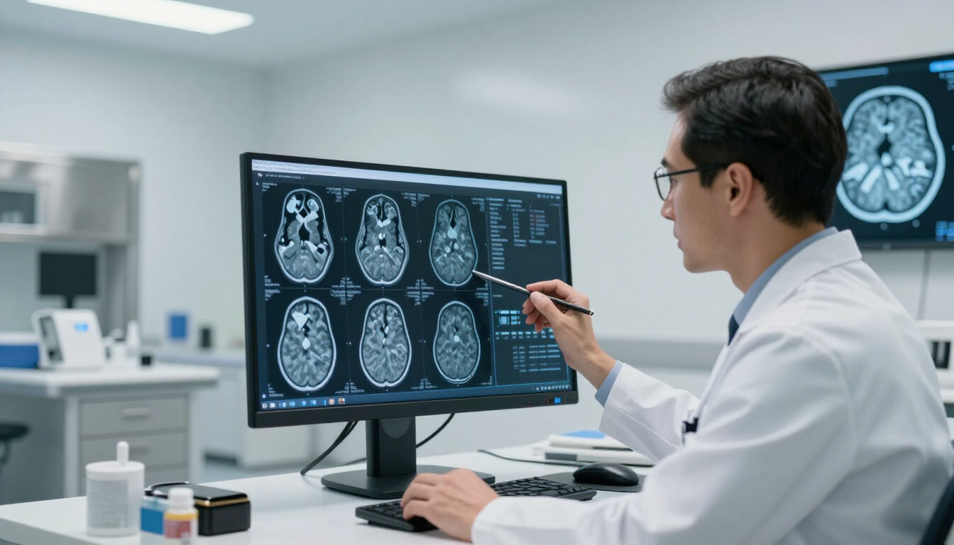
You might wonder why your doctor chose an MRI over a CT scan. Generally, an MRI is necessary when a higher level of detail is required for soft tissues. While a CT scan is excellent for looking at bones or detecting acute bleeding in an emergency, it lacks the resolution needed to see the subtle changes in brain architecture that indicate conditions like MS or early-stage dementia.
Doctors often recommend a cranial scan if you exhibit "red flag" symptoms. These aren't always signs of something serious, but they require professional investigation to ensure peace of mind and accurate diagnosis. For more health tips and medical news, feel free to visit MRI Plus News to stay informed about the latest in diagnostic technology.
Here are five specific scenarios where an MRI is the preferred choice:
- Multiple Sclerosis (MS): Detecting lesions or "plaques" in the brain and spinal cord.
- Epilepsy: Identifying structural causes for seizures, such as focal cortical dysplasia.
- Hormonal Issues: Examining the pituitary gland if hormone levels are abnormal.
- Chronic Vertigo: Checking the inner ear and auditory nerves for growths.
- Cognitive Decline: Ruling out treatable causes of memory loss or confusion.
How should I prepare for my scan?
Preparation for a brain MRI is relatively simple, but there are a few strict rules you must follow for safety. Because the machine uses a powerful magnet, the most important rule is the removal of all metallic objects. This includes jewellery, watches, piercings, and even some types of makeup that may contain metallic particles.
If you have any internal medical devices, you must inform the clinic immediately. While many modern implants are "MRI-safe," older pacemakers, cochlear implants, or certain aneurysm clips can be dangerous if placed inside a magnetic field.
In terms of lifestyle, you can usually eat and drink normally unless you are having a specific type of scan that requires fasting (which is rare for a standard brain MRI). If your scan requires a "contrast agent" to make certain structures show up more clearly, you may be asked to avoid heavy meals for an hour or two beforehand to prevent nausea.
What happens during a head MRI?
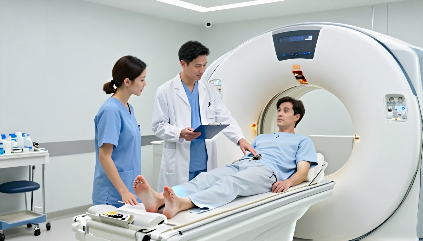
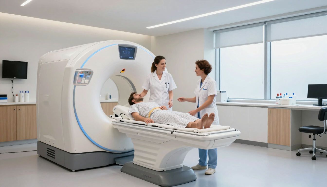
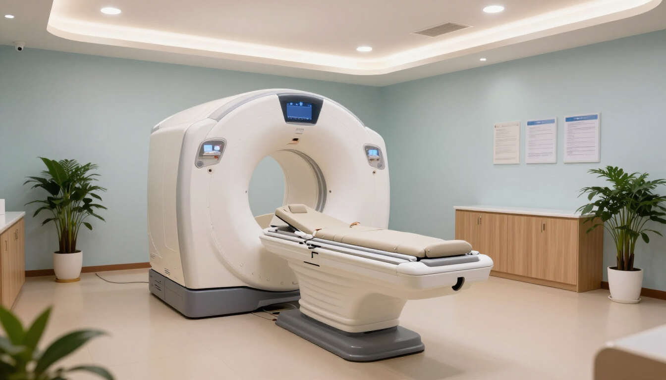
The experience of having a brain MRI is unique but straightforward. After changing into a gown and removing all metal, you will be asked to lie down on a motorized table. A special device called a "coil" will be placed around your head. This acts like an antenna to help capture the clearest possible images.
Once you are comfortable, the table will slide into the "bore" of the machine. The scanner is a large, tube-like structure. Throughout the procedure, which usually lasts between 20 and 45 minutes, you will hear a series of loud thumping or tapping noises. These are perfectly normal—they are the sounds of the magnetic coils shifting to create the imaging pulses.
To help you through the process, the clinical team will:
- Provide you with earplugs or headphones to dampen the noise.
- Give you a "call button" to hold so you can alert the staff at any time.
- Communicate with you through an intercom system between scan sequences.
- Allow you to listen to music or a radio station of your choice in some cases.
- Ensure you remain as still as possible to prevent blurring the images.
Managing anxiety and claustrophobia
It is completely normal to feel a bit nervous about being in a confined space. If you suffer from claustrophobia, it is best to discuss this with the provider beforehand. Many modern clinics, like MRI Plus, use "short-bore" or wider machines that feel much more open.
Conversational support from the radiographer can also make a huge difference. Knowing that you are being watched and that the scan can be paused if you feel overwhelmed provides a sense of control. Some patients find that closing their eyes before they slide into the machine and keeping them closed throughout the session helps them stay calm. Deep breathing exercises are also highly effective at lowering the heart rate during the louder parts of the scan.
How long does it take to get results?
If any issues appear in your scan images, our medical team will contact you to discuss them. Your imaging report will be sent to you electronically. You can use it for any follow-up consultations you may have. We can also offer copies of your images. Your report should be ready in 2 to 7 working days, depending on the package. We will contact your GP only in a medical emergency.
Common things a radiologist looks for include:
- Evidence of restricted blood flow (stroke).
- Abnormal growths or fluid collections (tumours/abscesses).
- Width of the brain's ventricles (checking for hydrocephalus).
- Integrity of the blood vessels (aneurysm screening).
- Signs of brain shrinkage or atrophy (dementia).
Why choose MRI Plus for your scan?
When it comes to your health, time is of the essence. Waiting months for a diagnostic scan can lead to unnecessary anxiety and delayed treatment. MRI Plus offers a faster, more accessible alternative. Based in London and Leigh on Sea, we focus on providing a seamless experience from booking to results.
We understand that medical procedures can be intimidating, which is why we emphasize transparency and clinical excellence. Our facilities are equipped with modern technology, and our staff are experts in making patients feel at ease. By choosing a private diagnostic provider, you gain control over your healthcare timeline.
Five reasons to choose private diagnostics:
- Fast Appointments Avilable: Secure a slot within days, not months.
- Expert Clinicians: Scans are reported by highly qualified Consultant Radiologists.
- Best Facilities: Benefit from clean, comfortable, and advanced clinical settings.
- Flexible Online Scheduling: Appointments that fit around your work and life.
- Fast Reporting: Get your results quickly within 2 days to move forward with your treatment plan.
Summary and Key Takeaways
A brain and head MRI is a safe, detailed way to look inside the skull. It can help diagnose many neurological issues. Whether you are dealing with chronic migraines, investigating a potential stroke, or monitoring a known condition, this scan provides the clarity needed for accurate medical decisions.
While the process involves lying still in a noisy machine, modern clinical practices ensure that patient comfort is a top priority. From ear protection to constant communication with the staff, the experience is designed to be as stress-free as possible. For those looking for the fastest route to answers, private providers like MRI Plus offer a streamlined, high-quality alternative to traditional healthcare pathways.
Key takeaways to remember:
- Safety First: MRI uses magnets, not radiation, making it very safe for most people.
- Detail Matters: It is the superior choice for soft tissue and neurological diagnosis.
- Preparation is Key: Always disclose any metallic implants or claustrophobia before the scan.
- Speed is Possible: Private diagnostics can significantly reduce your waiting time.
- Results are Comprehensive: A specialist radiologist provides a deep-dive report into your brain health.
If you have questions about your upcoming scan or wish to learn more about our services, visit our website, MRI Plus for more resources and information. Stay proactive about your health and get the answers you need today. Moving forward with a diagnosis is the first step toward effective treatment and long-term peace of mind.
References :
UHS. (2022). Patient information factsheet. In www.uhs.nhs.uk. https://www.uhs.nhs.uk/Media/UHS-website-2019/Patientinformation/Scansandx-rays/MRI-scans/Magnetic-resonance-imaging-MRI-brain-scan-2249-PIL.pdf
Website, N. (2025, September 12). MRI scan. nhs.uk. https://www.nhs.uk/tests-and-treatments/mri-scan/