
MRI of Hip: What to Expect and What It Shows
An MRI of the hip is a non-invasive diagnostic scan using magnetic fields to produce detailed images of bones, cartilage, ligaments, and tendons. It is the gold standard for identifying labral tears, stress fractures, and inflammatory conditions that standard X-rays often miss in active adults and athletes.
What does a hip MRI show?
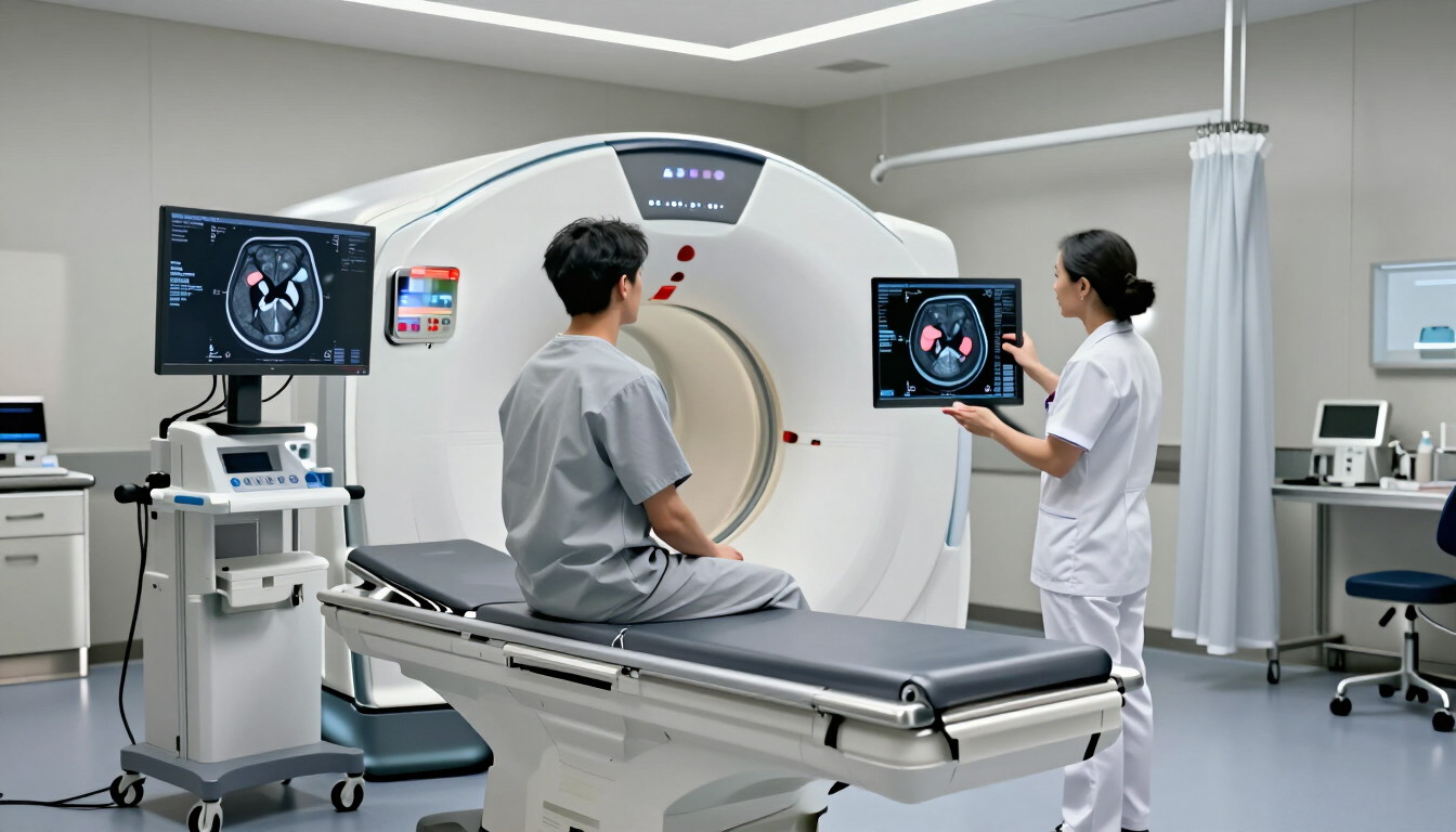
A hip MRI provides an incredibly detailed look at the internal structures of your joint, going far beyond what a standard X-ray can reveal. While X-rays are excellent for looking at significant bone breaks or advanced arthritis, an MRI is designed to see the "soft tissues" and the subtle changes within the bone itself. For an active person, these details are often the difference between a quick recovery and a chronic problem.
When you undergo an MRI of the hip, the radiologist is looking for several specific conditions:
- Labral Tears: The labrum is a ring of cartilage that follows the outside rim of your hip joint socket. Tears here are common in athletes and cause a clicking or catching sensation.
- Bursitis and Tendonitis: Inflammation of the small fluid-filled sacs (bursae) or the tendons that connect muscle to bone can be clearly identified.
- Bone Marrow Edema: This refers to swelling inside the bone, often a precursor to a stress fracture that wouldn't show up on other scans.
- Avascular Necrosis: A serious condition where the blood supply to the bone is interrupted, causing bone tissue to die.
- Cartilage Loss: Early signs of osteoarthritis can be spotted by looking at the thickness and health of the articular cartilage.
Understanding these structures helps your consultant create a targeted treatment plan. For younger patients, identifying a labral tear early can prevent long-term joint degradation. If you are experiencing groin pain that doesn't resolve with rest, this scan is usually the next logical step to find the root cause.
Does your whole body go in for a hip MRI?
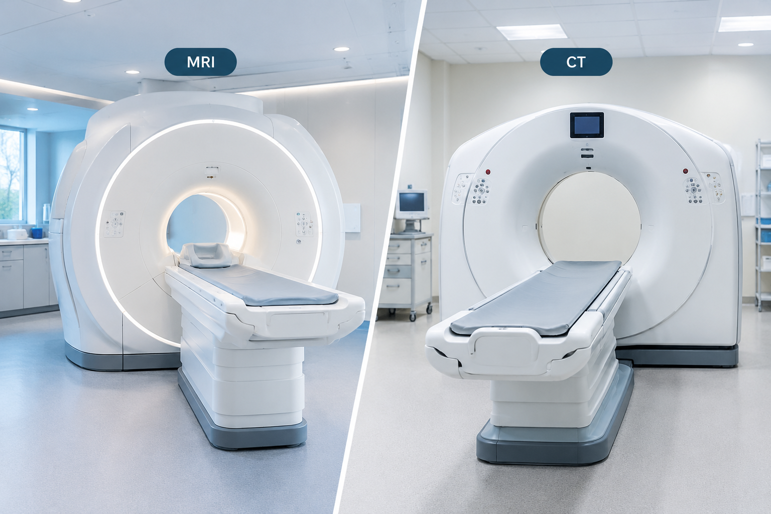
One of the most common concerns for patients is whether they will be fully enclosed in the "tunnel" or bore of the MRI machine. The short answer is: it depends on the specific machine and your height, but for a hip MRI, your head is often very close to the edge or even completely outside the scanner. Because the hip is located in the middle of your body, you will be positioned so that the hip joint is at the center of the magnetic field.
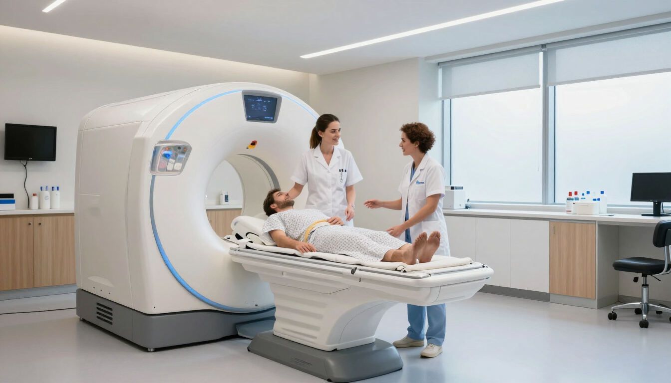
During the procedure, you will lie flat on a motorized table. A specialized piece of equipment called a "coil" is placed over your hip area. This act as an antenna to capture the radiofrequency signals from your body. The table then slides into the bore. For most modern
FAQs for HIP MRI :
Can a hip MRI show cancer?
Yes, a hip MRI can sometimes detect cancer, but it is not primarily performed as a cancer screening test.
Does hip MRI show pelvis?
Yes, a hip MRI can show parts of the pelvis, but its main focus is the hip joint and the surrounding structures.
How much does a private hip MRI cost in the UK?
A private hip MRI scan in the UK typically costs between £199 and £500, depending on the provider, location, and service level. At MRI Plus, a standard one-part hip MRI starts from £199, offering a competitively priced private MRI option.
References :
MRI Plus. Hip MRI Scan from £199. Available at: https://mriplus.co.uk/body-parts/hip
Radiological Society of North America (RSNA) and American College of Radiology (ACR). (n.d.-d). Hip MRI. Radiologyinfo.org. https://www.radiologyinfo.org/en/info/mri-hip