Does an MRI show nerve damage?
Yes, an MRI can show nerve damage by identifying physical changes like compression, inflammation, or structural irregularities. It mostly sees the cause ( a herniated disc for example ) . There are special methods such as MR Neurography that look specifically at the nerves to find the damage with high precision .
Living with unexplained pain, tingling, or numbness can be incredibly frustrating. When your body starts sending these signals, it is often a sign that your nervous system is under stress. Naturally, you want answers fast. In the world of modern diagnostics, the Magnetic Resonance Imaging (MRI) scan is one of the most powerful tools available to clinicians to peek inside the body without a single incision. But many patients ask: "Does an MRI actually show the nerve damage itself?"
Understanding how nerves appear on a scan is the first step toward getting the right treatment. Whether you are dealing with chronic back pain or a mysterious weakness in your hand, knowing what an MRI can—and cannot—see is vital for your healthcare journey.
How does an MRI detect nerve damage?
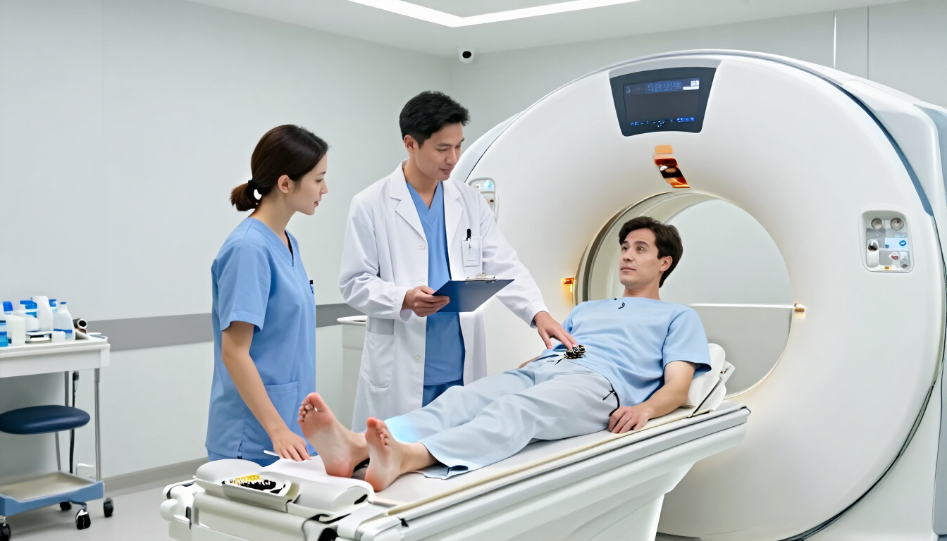
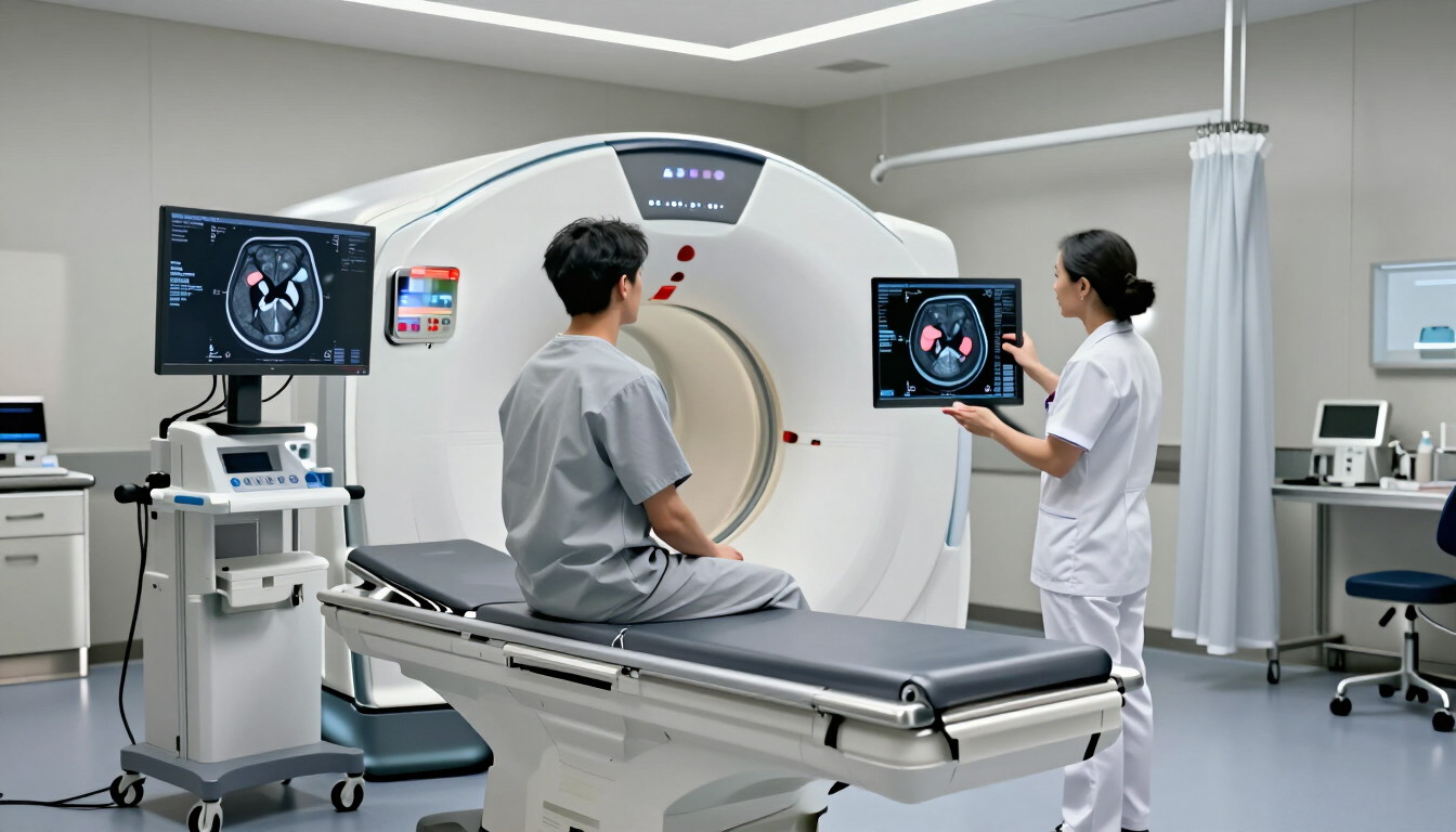
An MRI doesn’t just take a simple picture; it uses powerful magnets and radio waves to create highly detailed images of soft tissues. Nerves are considered soft tissues, but they are incredibly small and delicate. Standard MRI scans are excellent at showing the "environment" around a nerve. If a nerve is being pinched, squashed, or displaced, the MRI will show the culprit.
Common physical indicators of nerve issues on an MRI include:
- Compression: Seeing a disc or bone spur pressing directly against a nerve root.
- Inflammation: Changes in the signal intensity around a nerve can indicate swelling.
- Structural Changes: Identifying if a nerve has been physically torn or stretched.
- Displacement: Showing a nerve that has been pushed out of its natural pathway.
- Pathological Growths: Detecting tumors or cysts that are encroaching on neural space.
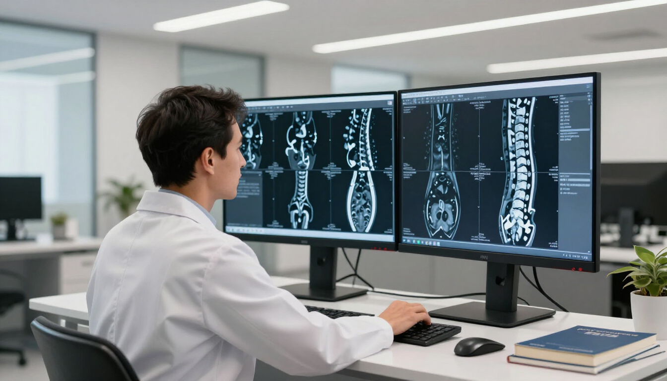
While a standard MRI is great for these structural issues, doctors sometimes use a specialized protocol called MR Neurography (MRN). This specific type of MRI uses different software settings to make the nerves "light up" or stand out from the surrounding muscle and fat. This allows radiologists to see the internal health of the nerve more clearly than a traditional scan might allow.
Which body parts are most common for nerve MRIs?
Nerve issues can happen anywhere, but certain areas of the body are more prone to compression and injury. At MRI Plus, we frequently see patients requiring investigations for specific problem areas, including the lumbar spine (sciatica and disc herniation), the cervical spine (pinched nerves causing arm numbness), the wrist (carpal tunnel syndrome), and the elbow (cubital tunnel syndrome). For more complex or diffuse nerve symptoms, we also offer dedicated brachial plexus scans, which image the nerve network supplying the arm and hand in high detail.
References :
Sneag, D. B., MD. (2022, January 15). MR Neurography (Magnetic Resonance Neurography). Hospital for Special Surgery. https://www.hss.edu/conditions_magnetic-resonance-neurography.asp
Barzouhi, A. E., Vleggeert-Lankamp, C. L. a. M., Nijeholt, G. J. L. À., Van Der Kallen, B. F., Van Den Hout, W. B., Verwoerd, A. J. H., Koes, B. W., Peul, W. C., & Group, F. T. L. H. S. I. P. S. (2013). Magnetic Resonance Imaging Interpretation in Patients with Sciatica Who Are Potential Candidates for Lumbar Disc Surgery. PLoS ONE, 8(7), e68411.
https://doi.org/10.1371/journal.pone.0068411