MRI of Knee: 5 Things to Know Before Your Scan
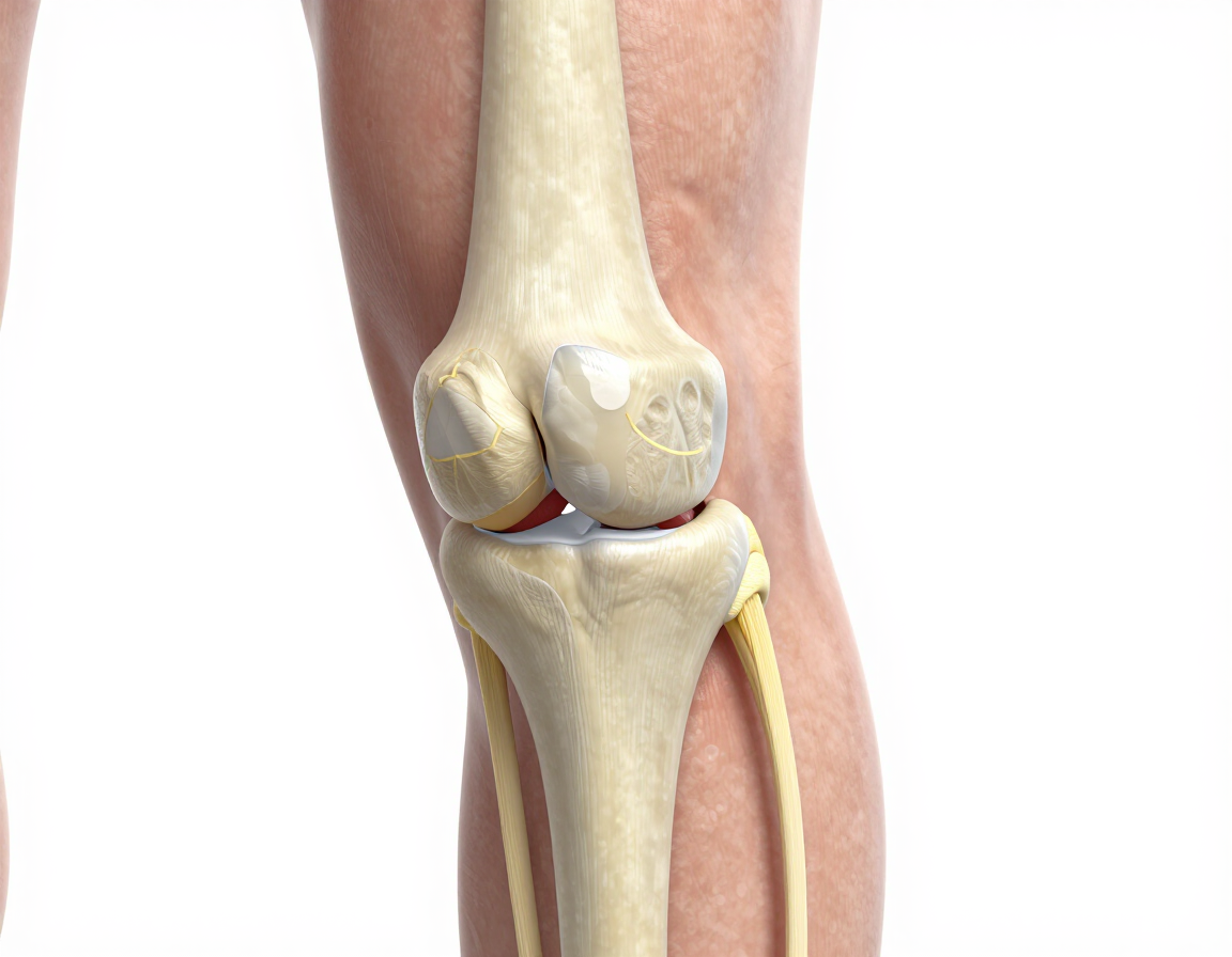
An MRI of the knee is a non-invasive diagnostic scan that uses powerful magnets and radio waves to create detailed images of the joint’s bones, cartilage, tendons, and ligaments. It is the gold standard for identifying ACL tears, meniscus injuries, and arthritis, helping doctors plan precise treatment.
What is an MRI of the knee used for?
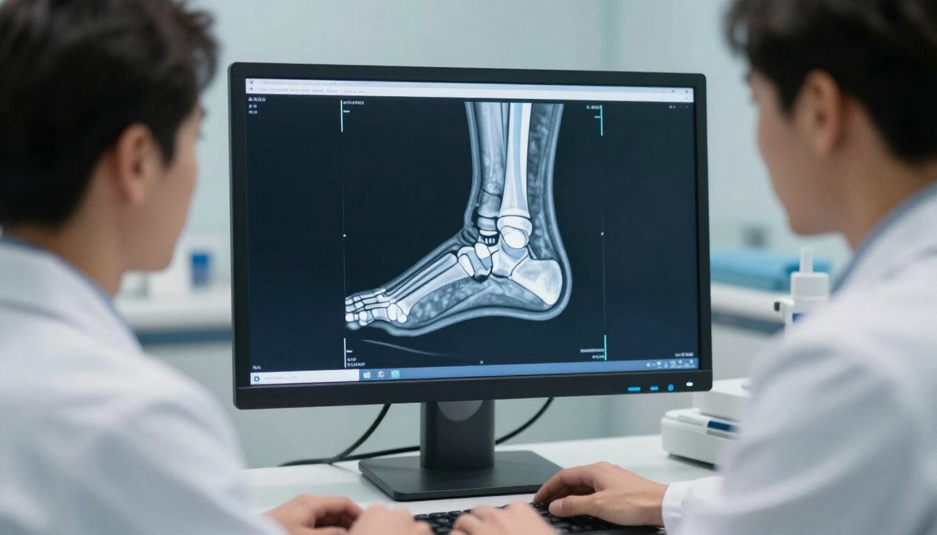
If you are experiencing persistent knee pain, swelling, or a limited range of motion, a medical professional will likely recommend an MRI of the knee. Unlike a standard X-ray, which primarily visualizes bone structures, an MRI provides a comprehensive view of the soft tissues. This makes it an essential tool for diagnosing complex internal injuries that might otherwise go unnoticed.
Common reasons for a knee MRI include:
- Investigating suspected ligament tears (such as the ACL or MCL).
- Checking for damage to the meniscus (the shock-absorbing cartilage).
- Evaluating unexplained knee pain or swelling that hasn't improved with rest.
- Assessing the extent of bone bruising or occult fractures.
- Monitoring the progression of degenerative conditions like osteoarthritis.
By providing a high-resolution map of the knee, this scan allows clinicians to decide whether a patient requires surgery, physical therapy, or a simple change in activity levels. For many athletes and active individuals, getting an accurate diagnosis quickly is the first step toward getting back on their feet.
How does the technology actually work?
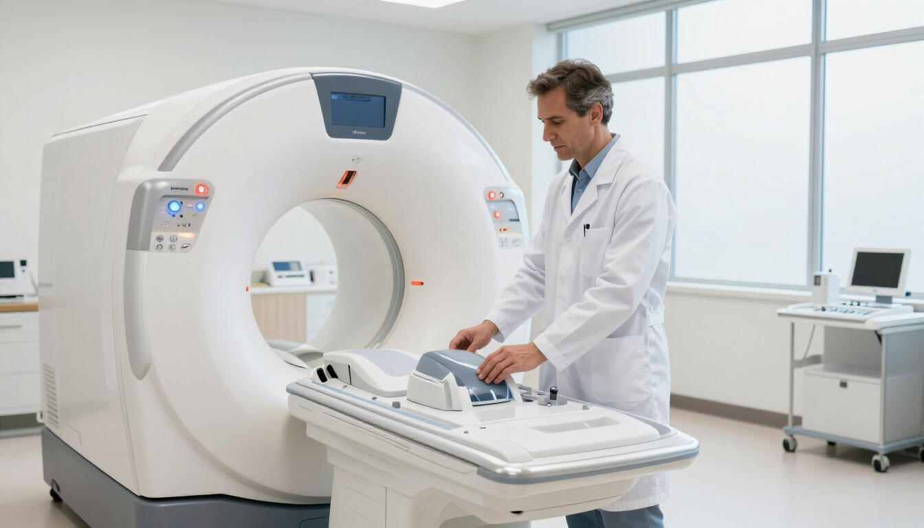
MRI stands for Magnetic Resonance Imaging. It is a marvel of modern physics that does not use ionizing radiation (unlike X-rays or CT scans). Instead, the machine creates a strong magnetic field that causes the protons in your body’s water molecules to align. When radio waves are pulsed through the area, these protons emit signals that a computer translates into 3D cross-sectional images.
When imaging the knee, the scan focuses on different "sequences." Some sequences are better at showing fluid (which indicates inflammation or swelling), while others are better at showing the fine structure of tendons. This multi-layered approach ensures that even the smallest micro-tears in the cartilage are visible to the radiologist.
Preparing for your appointment
Preparation for an MRI of the knee is generally very straightforward. Because the machine uses a powerful magnet, the most important step is ensuring you are not carrying or wearing any metallic objects. Most patients find the process simple and stress-free when they know what to expect.
Here are five key preparation tips:
- Wear comfortable clothing: Opt for loose-fitting clothes without metal zips, buttons, or underwires. Many clinics provide a gown if your clothing isn't suitable.
- Disclose medical implants: Always inform the staff if you have a pacemaker, cochlear implants, or any metal fragments in your body from previous surgeries or accidents.
- No fasting required: In most cases, you can eat, drink, and take your regular medications as usual before a knee MRI.
- Arrive early: Aim to arrive 15 minutes before your slot to complete the safety screening questionnaire.
- Bring previous scans: If you have had X-rays or older MRIs of the same knee, bringing the reports can help the radiologist compare changes over time.
If you suffer from claustrophobia, it is helpful to mention this when booking. Modern MRI machines are often wider than older models, and because only your lower body usually enters the center of the magnet for a knee scan, many patients find it much easier than a full-body or head scan.
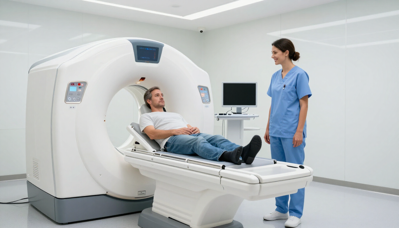
What to expect during the scan
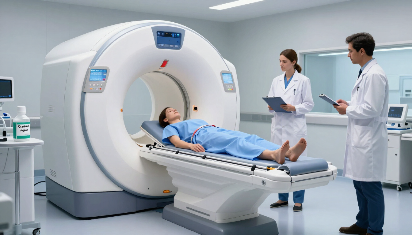
When you enter the scanning room, a radiographer will help you get positioned on the motorized table. Your knee will be placed inside a specialized device called a "coil." This coil acts like an antenna, helping the machine capture the clearest possible images of the joint.
The table will then slide into the large, tunnel-shaped scanner. For an MRI of the knee, your head will typically remain outside or near the edge of the tunnel, which significantly reduces any feeling of confinement. The most important thing you can do during the procedure is remain perfectly still. Even slight movements can blur the images, potentially requiring the radiographer to restart a sequence.
The machine is quite loud, making rhythmic thumping or humming noises. Most clinics provide earplugs or headphones so you can listen to music during the process. You will also be given a call button to hold, allowing you to speak to the radiographer at any time if you feel uncomfortable.
How long does an MRI of the knee take?
A standard MRI of the knee typically takes between 20 and 45 minutes . The exact duration depends on the complexity of the issue being investigated and whether your doctor has requested a "contrast" scan. A contrast scan involves a special dye being injected into your vein to highlight specific areas of inflammation or blood flow, which may add a bit of time to the appointment.
Compared to the weeks or months of waiting often found in traditional healthcare pathways, the actual time spent in the machine is quite short. At MRI Plus , we understand that your time is valuable. That is why we focus on streamlining every part of the process—from the moment you book to the delivery of your final report.
Common knee injuries detected by MRI
Because the knee is one of the most stressed joints in the human body, it is prone to a variety of injuries. An MRI is the most effective way to see exactly what is happening beneath the surface. Here are the most frequent findings:
- ACL (Anterior Cruciate Ligament) Tears: A common sports injury often caused by sudden stops or changes in direction.
- Meniscal Tears: Damage to the C-shaped cushions of cartilage between your shinbone and thighbone.
- Patellar Tendonitis: Inflammation of the tendon that connects your kneecap to your shinbone, often called "jumper’s knee."
- Bone Marrow Edema: Swelling inside the bone itself, which can be a sign of a stress fracture or severe bruising.
- Bursitis: Inflammation of the small, fluid-filled sacs that cushion the knee joint.
By identifying these issues early, you can avoid further damage. For instance, continuing to walk on a torn meniscus without knowing it can lead to accelerated wear and tear of the joint, eventually resulting in early-onset arthritis.
Why should you choose a private knee MRI?
Choosing a private diagnostic provider like MRI Plus offers several distinct advantages over traditional public routes. The primary factor for most patients is speed . In healthcare, time is often the most critical factor in recovery. The longer an injury goes undiagnosed, the more likely it is that compensatory movements will lead to pain in other areas, such as the hips or lower back.
Benefits of opting for a private scan include:
- Rapid Appointments: Often available within days rather than months.
- Flexible Scheduling: Evening and weekend slots to suit a busy lifestyle.
- Fast Reporting: Clinical results are typically delivered to you or your consultant within 24 to 48 hours.
- Peace of Mind: Eliminating the stress of the unknown allows you to start the correct treatment immediately.
Our approach at MRI Plus is centered on clinical quality and transparency. We believe that everyone should have access to high-end diagnostics without the friction of long waiting lists. Whether you are a professional athlete or someone who just wants to enjoy a walk in the park without pain, fast diagnostics are key.
Understanding your results and next steps
Once your MRI of the knee is complete, the images are sent to a consultant radiologist—a doctor who specializes in interpreting medical scans. They will write a detailed report describing the state of your bones, cartilage, and ligaments.
It is important to remember that the MRI report is just one piece of the puzzle. Your GP or orthopedic consultant will use this report alongside a physical examination to create your treatment plan. This might include:
- Physiotherapy: To strengthen the muscles supporting the knee.
- Injections: Such as corticosteroids or hyaluronic acid to reduce inflammation.
- Surgery: If the scan shows a significant tear that cannot heal on its own.
- Conservative Management: Rest, ice, and lifestyle adjustments.
Receiving your results quickly means you can have an informed conversation with your doctor sooner, moving you one step closer to recovery.
Summary of MRI Knee Insights
An MRI of the knee is the most reliable way to diagnose internal joint issues and get your health back on track. By choosing a fast, efficient provider, you can bypass long waiting times and gain immediate clarity on your condition.
- Immediate Answers: MRI provides the most detailed view of soft tissue injuries like ACL and meniscus tears.
- No Radiation: It is a safe, painless procedure using magnetic technology.
- Preparation is Easy: Simply wear metal-free clothing and stay still during the 20-40 minute scan.
- Faster Recovery: Quick diagnostics allow for earlier intervention and better long-term outcomes.
- Take Action: If you have persistent knee pain, consult a professional about booking a scan today.
References:
Knee MRI for suspected meniscal tears - EBI. (2024, September 25). EBI. https://ebi.aomrc.org.uk/interventions/knee-mri-for-suspected-meniscal-tears/
NHS Scotland. MRI Criteria – Knee. Available at: https://www.rightdecisions.scot.nhs.uk/
Knee MRI Scan from £199 | MRI Plus. (n.d.). https://mriplus.co.uk/body-parts/knee