Small Intestine MRI: Procedure Guide
A Small Intestine MRI , also known as MRI Enterography, is a non-invasive medical imaging procedure used to produce detailed pictures of the small bowel. It is specifically designed to identify inflammation, bleeding, tumors, and conditions like Crohn’s disease by using magnetic fields and radio waves rather than harmful ionizing radiation.
If you have been experiencing persistent abdominal pain, unexplained weight loss, or digestive issues, your doctor may recommend this specialized scan. Unlike a standard abdominal MRI, this procedure focuses specifically on the miles of tubing that make up your small intestine, providing a level of detail that other tests might miss.
What can a small intestine MRI detect?
The small intestine is one of the more difficult areas of the body to visualize. Because it is so long and folded, traditional methods like endoscopy or colonoscopy often can’t reach every part of it. This is where the small bowel MRI comes in. It is an incredibly powerful tool for diagnosing a range of complex conditions.
One of the most common reasons patients are referred for this scan is to evaluate Crohn’s disease. It allows clinicians to see not just the surface of the bowel, but also the thickness of the bowel wall and any complications like fistulas or abscesses. Beyond inflammatory conditions, this scan is vital for spotting small tumors or polyps that could be causing a blockage.
It is also frequently used to investigate the source of unexplained gastrointestinal bleeding. If other tests have come back clear but you are still showing signs of blood loss, the detailed imaging of a small bowel MRI can often pinpoint the exact location of the issue. Furthermore, it can identify structural problems like strictures (narrowing of the bowel) or obstructions that might be causing you significant discomfort.
Understanding the MRI Plus approach to diagnostics
At MRI Plus, we understand that waiting for medical answers can be a stressful time. That is why we have built our service around speed and clinical accuracy. While many traditional healthcare pathways involve long waiting lists and delays, we focus on getting you into the scanner and getting your results back to your consultant as quickly as possible.
We combine state-of-the-art technology with a patient-first philosophy. This means that from the moment you book your appointment to the time you receive your digital report, every step is designed to be smooth and transparent. Our goal is to provide the clinical clarity you need to move forward with your treatment plan, ensuring that no symptom goes uninvestigated for longer than necessary. Our expertise in MRI Plus and diagnostic pathways ensures that you are in safe hands throughout the entire journey.
How should I prepare for a small intestine MRI?
Preparation is key to getting the clearest possible images. Because the small intestine is usually collapsed when empty, we need to "distend" or expand the bowel so the radiologist can see the walls clearly. To do this, you will usually be asked to follow specific dietary instructions in the 24 hours leading up to your scan.
Typically, you will need to fast for several hours before your appointment—usually around 4 to 6 hours. This ensures that food particles don't interfere with the images. However, the most important part of the preparation happens right before the scan begins. You will be asked to drink a specific amount of contrast liquid (often containing mannitol or sorbitol) over the course of about an hour. This liquid fills the small bowel, allowing it to show up clearly on the MRI.
Important preparation tips include:
- Wearing comfortable, loose-fitting clothing without metal zippers.
- Removing all jewelry, piercings, and watches before the scan.
- Informing the staff if you have any metal implants or pacemakers.
- Bringing a list of any medications you are currently taking.
- Staying hydrated before your fasting period begins.
What happens during the small intestine MRI procedure?
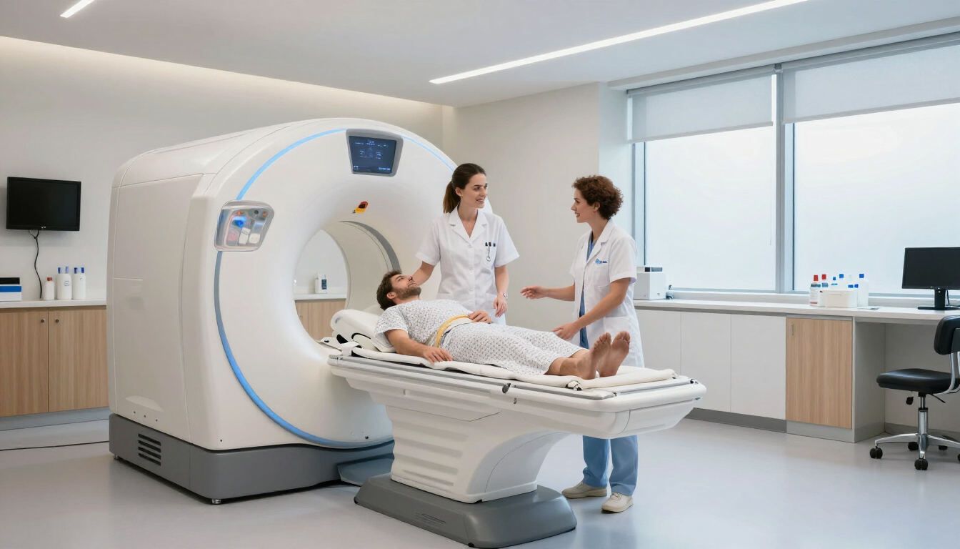
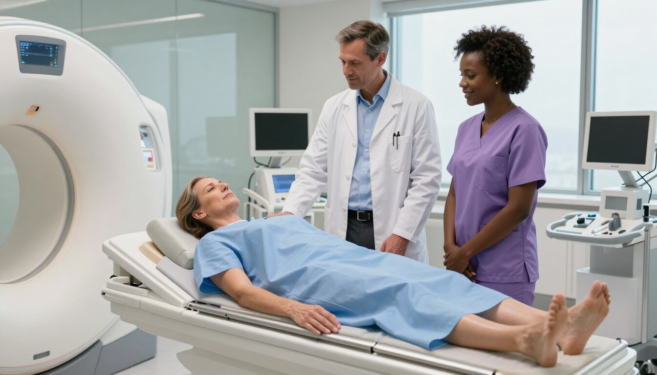
When you arrive for your Small Intestine MRI, the process is straightforward but requires a bit of patience. After you have finished drinking the contrast liquid, you will be guided to the MRI suite. You will lie on a motorized table that slides into the center of the MRI machine, which looks like a large, brightly lit tunnel.
During the scan, the radiographer will be in constant communication with you through an intercom. You will hear loud clicking or thumping sounds—this is perfectly normal and is just the machine’s magnets working to create the images. We often provide headphones so you can listen to music or the radio to help you relax. You will need to stay as still as possible, and at certain points, the radiographer may ask you to hold your breath for a few seconds to ensure the images aren't blurred by your breathing movements.
In some cases, an intravenous (IV) contrast agent called gadolinium may also be injected into your arm halfway through the procedure. This helps highlight blood flow and areas of inflammation even more clearly. The entire scanning process usually takes between 30 and 60 minutes, depending on the specific details your consultant needs.

Does a small intestine MRI hurt?
The procedure itself is entirely painless. There are no needles involved in the imaging itself, other than the optional IV contrast injection which feels like a small pinch. Some patients find the drinking of the contrast liquid a bit tedious, as it involves consuming a large volume in a short time, and it may cause some mild bloating or a frequent need to visit the bathroom afterward.
However, the MRI technology is non-invasive and does not use radiation, making it a very safe option for most people. If you feel anxious about enclosed spaces, please let our team know beforehand. We are experienced in helping claustrophobic patients feel comfortable and can offer various techniques to help you through the process.
Benefits of MRI over other imaging types
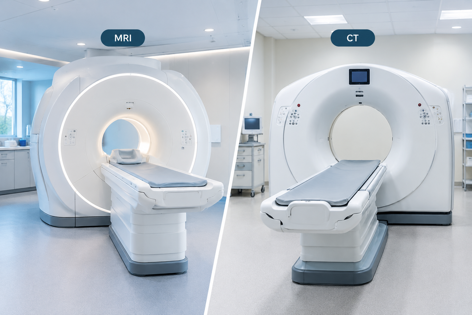
You might wonder why a Small Intestine MRI is preferred over a CT scan or an X-ray. The primary benefit is the lack of radiation. Because the small intestine often requires repeat monitoring—especially for chronic conditions like Crohn’s—avoiding cumulative radiation exposure is a significant health advantage.
Furthermore, MRI offers superior "soft tissue contrast." This means it is much better at distinguishing between different types of tissue, such as healthy bowel versus inflamed or scarred tissue. It provides a three-dimensional view that allows doctors to see the bowel from multiple angles, which is invaluable for surgical planning or monitoring how well a patient is responding to a new medication. By choosing MRI Plus, you are opting for a diagnostic tool that prioritizes both your safety and the precision of your results.
Fast results for better patient outcomes
Once the scan is complete, the images are sent to a specialist radiologist for review. They will analyze the hundreds of detailed pictures to look for any abnormalities. In the traditional system, getting these results back can sometimes take weeks. At MRI Plus, we prioritize a streamlined reporting process.
We know that your peace of mind depends on getting answers quickly. Our digital reporting system ensures that once the radiologist has completed their assessment, the findings are made available to your healthcare provider without unnecessary administrative delays. This speed allows you to start your treatment or move to the next stage of your diagnostic journey much sooner, reducing the time spent in a state of uncertainty.
Summary and Key Takeaways
A Small Intestine MRI is a vital, radiation-free diagnostic tool that provides deep insights into your digestive health. Whether you are managing a chronic condition or searching for the cause of new symptoms, this procedure offers the clarity needed for an accurate diagnosis.
Key takeaways for your procedure:
- The scan is primarily used for Crohn's disease, tumors, and unexplained pain.
- You will need to fast and drink a contrast liquid to prepare your bowel.
- The procedure is painless and takes approximately 45 to 60 minutes.
- MRI is safer than CT scans for long-term monitoring due to the lack of radiation.
- Choosing a private provider like MRI Plus ensures faster access and rapid reporting.
If you have further questions or wish to book an appointment, please visit our latest updates at MRI Plus News to see how we are continuing to lead the way in private diagnostic care. Your health and comfort are our top priorities, and we are here to support you every step of the way.
Reference :
Having an MRI enterography (small bowel scan). (2025). https://www.royalberkshire.nhs.uk/media/ufsf12r3/having-a-mri-enterography_jul25.pdf
Radiological Society of North America (RSNA) and American College of Radiology (ACR). (n.d.-e). MR enterography. Radiologyinfo.org. https://www.radiologyinfo.org/en/info/mrenterography
Small Bowel MRI Scan from £199 | MRI Plus. (n.d.).
https://mriplus.co.uk/body-parts/small-bowel