What Does a Small Bowel MRI Scan Show? (2026)
A small bowel MRI scan is a non-invasive examination that creates detailed images of the small intestine and surrounding tissues. It can show inflammation, narrowing, obstruction, fistulas, bleeding-related changes, abscesses, and some tumours. Doctors commonly use it to assess Crohn’s disease, inflammatory bowel disease (IBD), and unexplained gastrointestinal symptoms.
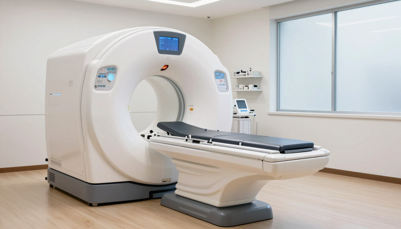
What does a small bowel MRI scan show?
A small bowel MRI scan, sometimes called MR enterography, produces detailed cross-sectional images without using X-rays. It helps clinicians examine the bowel wall, the space around the bowel, and nearby abdominal structures.
Depending on your symptoms and the reason for referral, the scan may help identify:
- Inflammation and bowel wall thickening linked with Crohn’s disease or other IBD
- Narrowed sections of bowel, known as strictures
- Partial or complete obstructions
- Fistulas, abscesses, and other complications of bowel inflammation
- Small bowel tumours or abnormal tissue
MRI can also show how the bowel moves and how substances pass through it. This can give your specialist useful information about disease activity and whether treatment is working.
Why might a doctor recommend a small bowel MRI?
Your doctor may recommend this examination if you have confirmed or suspected Crohn’s disease, inflammatory bowel disease, unexplained gastrointestinal bleeding, or symptoms suggesting a blockage. It may also be used when other tests have not provided enough information.
A small bowel MRI can help your healthcare team to:
- Assess the extent and severity of bowel disease
- Check for complications without exposing you to radiation
- Investigate persistent abdominal pain, diarrhoea, weight loss, or bleeding
- Monitor changes over time
- Plan treatment or decide whether further investigation is needed
For people who need repeated imaging, avoiding radiation can be particularly helpful. Your referring clinician will decide whether MRI is appropriate based on your medical history, symptoms, and previous test results.
How should you prepare for a small bowel MRI scan?
Preparation instructions can vary between clinics, so follow the guidance given with your appointment. In many cases, you will be asked to avoid solid food for a period before the scan while continuing to drink clear fluids as advised.
You may be asked to:
- Arrive early so the team can complete safety checks and explain the examination.
- Drink a specific amount of fluid shortly before the scan. This helps expand the small bowel so its walls are easier to assess.
- Tell the radiographer about medicines, allergies, kidney problems, previous operations, or possible pregnancy.
- Remove metal objects, including jewellery, watches, hair accessories, and some clothing items.
- Bring relevant referral letters, medication details, and previous scan information if requested.
Do not stop prescribed medication unless your healthcare professional tells you to. If you are unsure about eating, drinking, or taking medication before your appointment, contact the clinic for clarification.
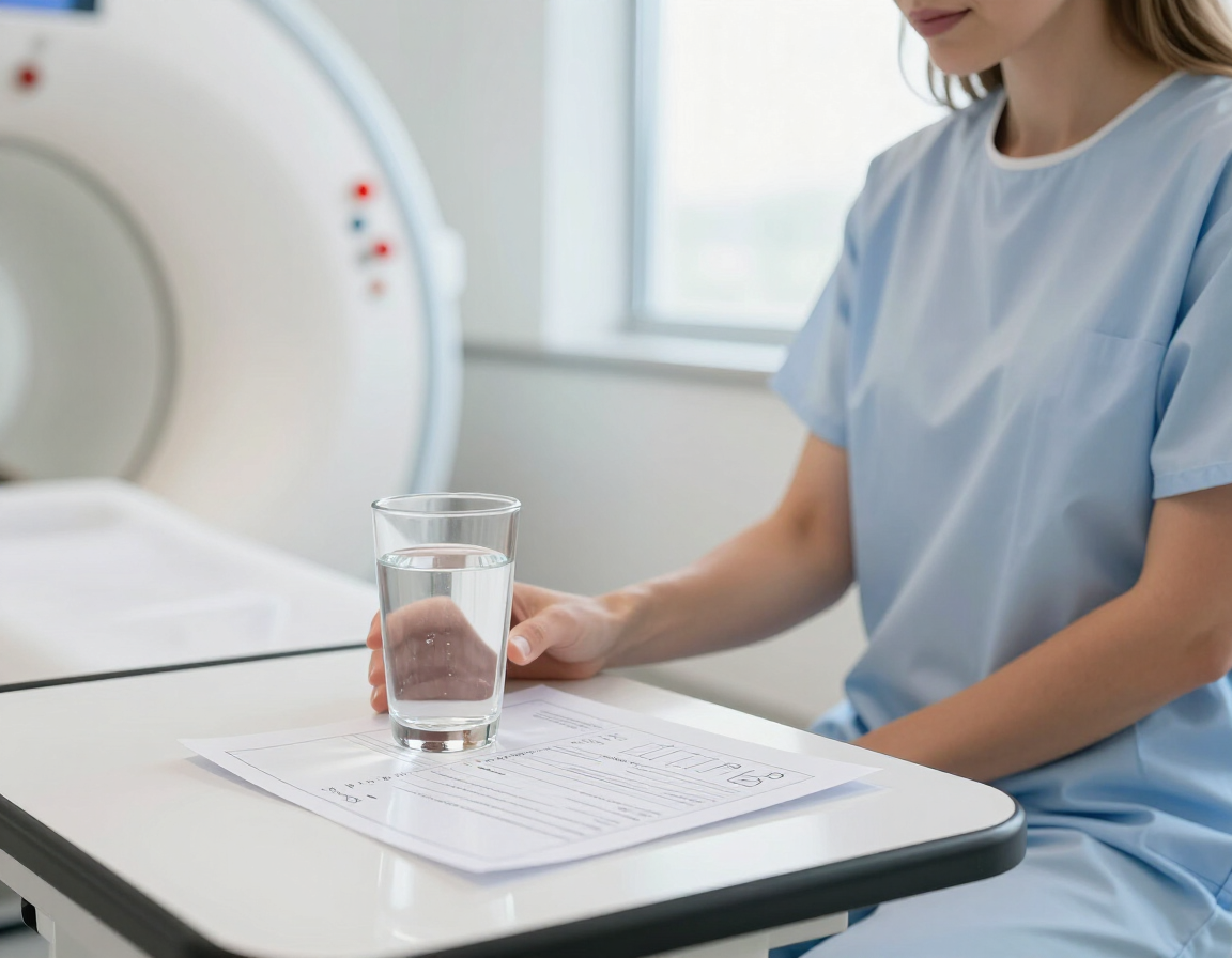
What happens during a small bowel MRI scan?
When you arrive, a radiographer will explain the process and ask safety questions. You will usually change if needed and remove anything that could interfere with the magnetic field.
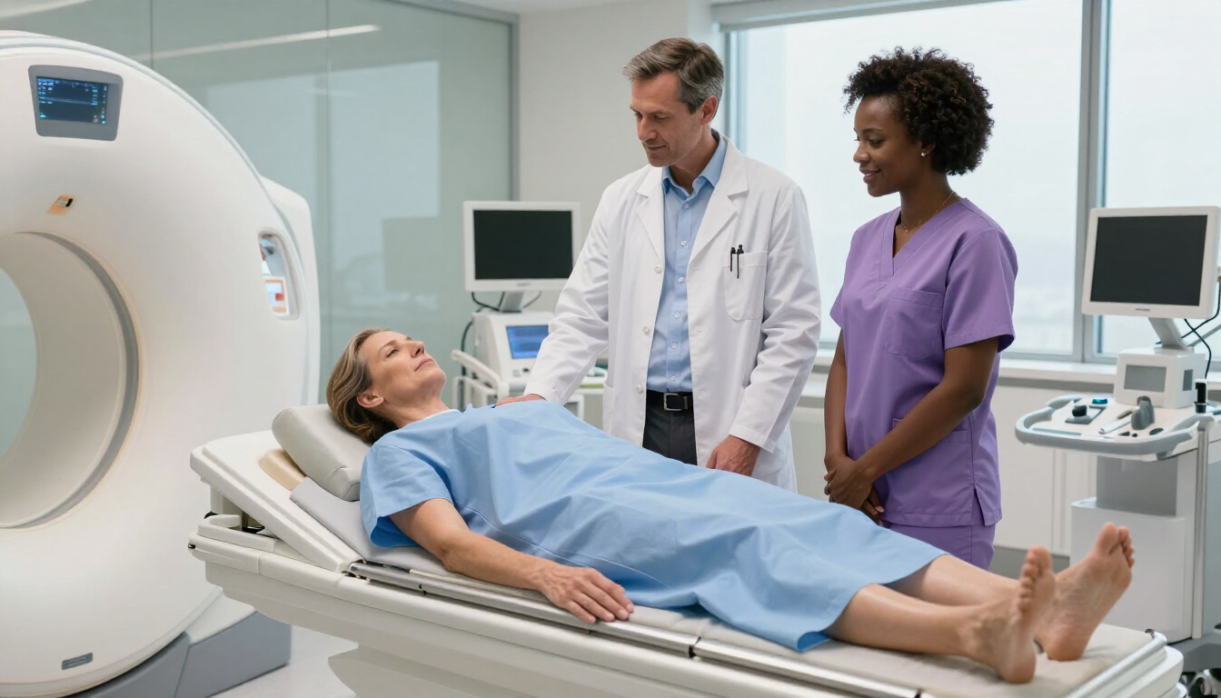
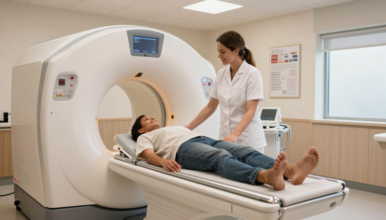
You will lie on a padded table that moves into the MRI scanner. The scanner is a short, open-ended tube. It does not use ionising radiation, but it makes loud tapping and knocking sounds. Earplugs or headphones are normally provided.
The radiographer may ask you to hold your breath for short periods while images are taken. Staying as still as possible helps produce clear pictures. You can communicate with the team throughout the examination, and an alert button is usually available if you need assistance.
Some small bowel MRI examinations involve an injection of contrast material. Contrast can improve the visibility of inflammation, blood vessels, and abnormal tissue. It is important to tell the team about kidney disease, previous reactions to contrast, or other relevant medical conditions.
The scan itself commonly takes around 30 to 60 minutes, although your total appointment may be longer because of preparation and post-scan checks.
Is a small bowel MRI scan uncomfortable?
Most people find the scan painless. The main challenges may be lying still, hearing the scanner sounds, or feeling enclosed. Drinking the preparation fluid can cause temporary fullness, bloating, or mild nausea in some patients.
If you feel anxious in enclosed spaces, tell the clinic before your appointment. The team may be able to discuss practical support or other options. Let the radiographer know immediately if you feel unwell or uncomfortable during the examination.
MRI does not expose you to ionising radiation. However, because it uses a strong magnetic field, you must disclose any implanted medical devices, metal fragments, clips, or previous surgery. Some devices are MRI-compatible, but they must be checked before scanning.
What happens after the scan?
After the examination, most people can return to normal activities straight away. If contrast was used, you may be advised to drink fluids unless your clinician has given you different instructions.
You could experience short-lived bloating or loose stools after drinking the bowel preparation. These effects usually settle. If you develop severe pain, persistent vomiting, breathing difficulty, swelling, or another worrying symptom, seek urgent medical advice.
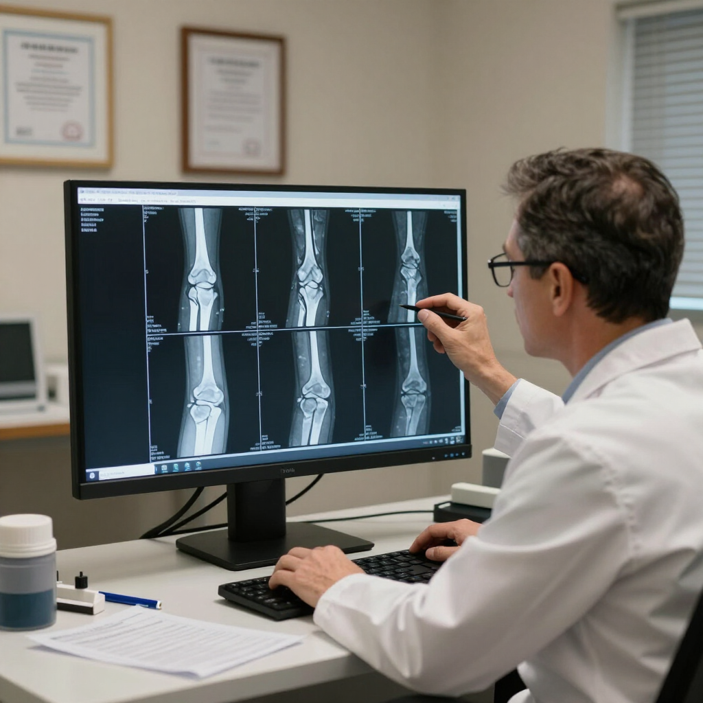
A specialist radiologist reviews the images and prepares a report for the referring clinician. Results are not always available immediately because the images need careful analysis. Ask your referrer or diagnostic provider when and how you will receive the report.
How accurate is a small bowel MRI?
Small bowel MRI can provide highly detailed information, particularly about bowel inflammation, strictures, fistulas, abscesses, and surrounding tissues. Its usefulness depends on image quality, preparation, the condition being assessed, and how the findings fit with your symptoms and other tests.
No single investigation answers every clinical question. Your doctor may interpret the MRI alongside blood tests, stool tests, endoscopy, ultrasound, CT, or biopsy results. A scan can identify an area that needs further assessment, but it does not always provide a final diagnosis on its own.
Small bowel MRI compared with other tests
The most suitable test depends on what your clinician needs to investigate.
- MRI: Detailed images of the bowel and surrounding tissues without ionising radiation; often useful for monitoring Crohn’s disease.
- CT: Fast and widely available, but uses ionising radiation; may be preferred in urgent situations.
- Colonoscopy: Allows direct examination and biopsy of the colon and the end of the small bowel, but does not view the entire small intestine.
- Capsule endoscopy: Uses a swallowed camera to view much of the small bowel, but may not be suitable where an obstruction or narrowing is suspected.
Your clinician will weigh the benefits, limitations, urgency, and your medical history before recommending a test.
Frequently asked questions
Can a small bowel MRI detect Crohn’s disease?
It can show features associated with Crohn’s disease, including inflammation, bowel wall thickening, strictures, fistulas, and abscesses. The diagnosis and treatment plan are based on the MRI report alongside symptoms, blood or stool tests, endoscopy, and clinical assessment.
Can MRI find a small bowel tumour?
MRI may identify an abnormal mass or changes in nearby tissues. If something unusual is seen, your specialist may recommend further imaging, endoscopy, biopsy, or another test to confirm what it is.
Can I have an MRI if I am claustrophobic?
Many people with claustrophobia complete an MRI successfully with support. Tell the clinic in advance so the team can discuss the scanner, communication methods, and possible adjustments. Never take a sedative unless it has been prescribed and arranged safely.
How long do small bowel MRI results take?
Timing varies by provider and clinical urgency. The radiologist must review the images and send a report to the referring clinician, who will explain the findings and next steps.
Key takeaways
A small bowel MRI scan gives detailed, radiation-free images of the small intestine and surrounding tissues. It is especially useful for assessing Crohn’s disease, IBD, strictures, obstruction, fistulas, abscesses, and some tumours. Prepare by following your clinic’s food, drink, and medication instructions, and tell the team about implants, kidney problems, allergies, or pregnancy. The scan is usually painless and takes around 30 to 60 minutes. If your clinician has recommended one, confirm the preparation requirements and ask when your results will be discussed. For further updates from MRI Plus, visit MRI Plus News .