What Does an Adrenal MRI Scan Show?
An adrenal MRI scan provides detailed images of the adrenal glands, helping clinicians assess an incidentaloma, determine whether a mass is benign or suspicious, and guide follow-up. It can show a lesion’s size, structure, fat, fluid, blood products and relationship to nearby tissues without using ionising radiation.

What is an adrenal incidentaloma?
An adrenal incidentaloma is a growth or mass on an adrenal gland that is found unexpectedly, often during a CT scan or MRI performed for another reason. The adrenal glands sit above the kidneys and produce hormones including cortisol, adrenaline and aldosterone.
Most incidentalomas are non-cancerous and do not cause symptoms. However, doctors may recommend further assessment to understand:
- Whether the mass has features suggesting a benign or malignant growth
- Whether it is producing excess hormones
- Whether it has changed in size over time
- Whether treatment or monitoring is appropriate
An unexpected finding can understandably cause concern. An adrenal MRI scan is one part of a wider assessment and is interpreted alongside your symptoms, medical history, blood tests and any previous imaging.
Why might you need an adrenal MRI scan?
Your doctor may recommend an adrenal MRI scan if an adrenal lesion needs more detailed characterisation. MRI can provide excellent soft-tissue contrast, which helps radiologists distinguish different tissue types within a mass.
An MRI may be suggested when:
- A previous scan did not clearly identify the type of lesion
- The mass has an indeterminate appearance
- A patient is younger or pregnant and avoiding radiation is preferable
- The clinician needs a closer look at the adrenal glands and surrounding structures
- Follow-up imaging is required to assess stability
MRI is not automatically needed for every incidentaloma. The appropriate scan depends on the lesion’s appearance, size, clinical context and the advice of your referring clinician.
What does an adrenal MRI scan show?
An adrenal MRI scan can show the location, size, shape and internal characteristics of an adrenal mass. Radiologists use several sequences to assess how the lesion behaves on different types of images.
The scan may help show:
- Fat: Certain patterns of fat can support a benign diagnosis, such as an adrenal adenoma or myelolipoma.
- Fluid: Cysts and some other lesions can have fluid-like characteristics.
- Internal structure: MRI can reveal whether a mass is uniform or contains areas of bleeding, degeneration or necrosis.
- Contrast behaviour: If contrast is used, enhancement patterns may provide additional information.
- Nearby anatomy: The scan can show the relationship between the adrenal gland, kidneys, blood vessels and surrounding tissues.
- Change over time: Comparison with earlier imaging can help establish whether a lesion is stable or growing.
MRI findings can be reassuring, but imaging alone does not always provide a final diagnosis. Your specialist may combine the report with hormone testing or recommend further review.

Can an MRI tell if an adrenal mass is cancerous?
An MRI can identify features that are more or less concerning, but it cannot guarantee whether every adrenal mass is cancerous. The radiologist assesses the lesion’s size, margins, tissue characteristics, enhancement and relationship with nearby structures.
A report may describe a mass as having characteristics typical of a benign lesion, indeterminate features or suspicious features. Suspicious findings do not automatically mean cancer; they indicate that additional assessment may be necessary.
Your care team might consider:
- Blood or urine tests for hormone production
- Comparison with previous scans
- Repeat imaging after an appropriate interval
- Referral to an endocrinologist, surgeon or specialist multidisciplinary team
- Additional imaging or treatment, depending on the overall assessment
Ask your referring clinician to explain the result in the context of your personal circumstances rather than interpreting a single phrase in the report alone.
Does an adrenal MRI check hormone production?
No. An adrenal MRI shows anatomy and tissue characteristics, but it does not determine by itself whether a mass is producing hormones. Functional assessment usually involves blood and/or urine tests requested by your doctor.
Hormone testing may be considered for cortisol, aldosterone, adrenaline-related hormones or other markers, depending on your symptoms and medical history. Symptoms such as unexplained high blood pressure, episodes of palpitations, sweating, headaches, easy bruising or changes in weight should be discussed with your healthcare professional.
A person can have a non-functioning incidentaloma, while a relatively small mass can sometimes produce hormones. This is why imaging and clinical testing are complementary rather than interchangeable.
What happens during an adrenal MRI scan?
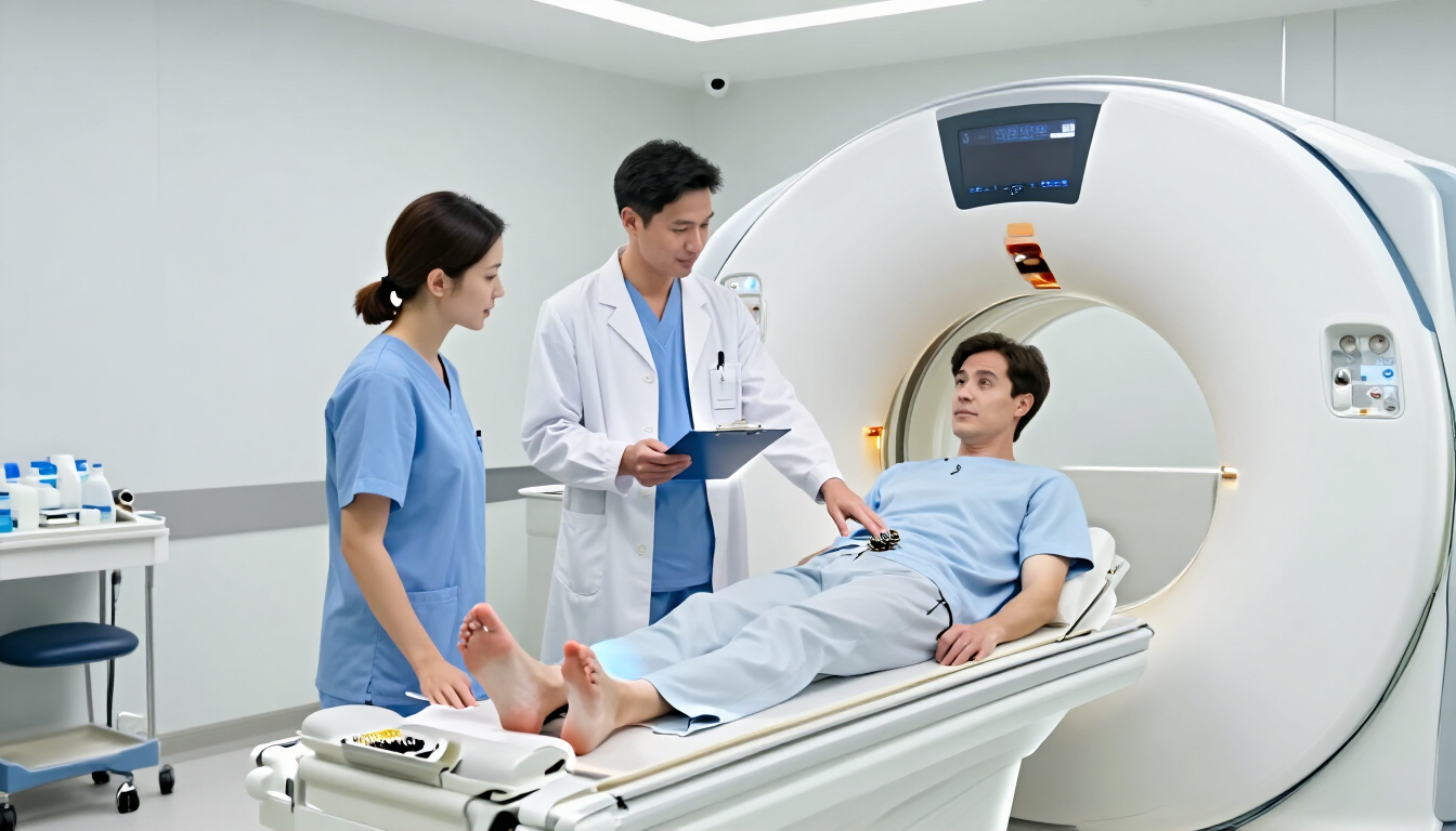
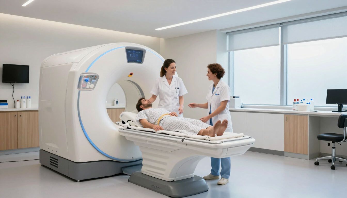
An adrenal MRI is usually performed while you lie on your back on a padded table that moves into the scanner. The examination is painless, although the machine makes loud tapping and knocking sounds. You may receive headphones or ear protection.
The radiographer may ask you to remain still and hold your breath briefly at different points. Depending on the referral and the type of lesion being assessed, contrast may be given through a small cannula in your arm.
Before the appointment, tell the imaging team if you:
- Have a pacemaker, implanted device, metal fragment or other medical implant
- Have kidney disease or a previous reaction to contrast
- Could be pregnant
- Feel anxious in enclosed spaces
- Need help with mobility or communication
Most adrenal MRI appointments take around 20 to 40 minutes, although timing varies. Follow the instructions provided by the imaging centre, including any guidance about eating, drinking or medicines.
Is contrast needed for an adrenal MRI?
Sometimes. The decision depends on the clinical question, the appearance of the lesion and the radiologist’s protocol. Contrast can help assess enhancement and improve characterisation, but many adrenal MRI examinations can provide useful information without it.
If contrast is proposed, the imaging team will check relevant medical information and explain the process. Tell them about kidney problems, allergies, previous contrast reactions and pregnancy. Do not stop prescribed medicines unless your clinician specifically advises you to do so.
How long do adrenal MRI results take?
After the scan, a radiologist reviews the images and prepares a report for the referring clinician. The report may comment on the adrenal glands, the size and appearance of any lesion, comparison with prior imaging and suggested follow-up.
The time to receive results depends on the provider and referral pathway. Your clinician or diagnostic centre should explain when and how results will be shared. If you are arranging a private scan, ask about reporting times, how the report is delivered and whether images can be shared with your GP or consultant.
For updates about diagnostic services and patient information, visit the our MRI Plus.

What should you ask after the scan?
When discussing your results, useful questions include:
- What type of adrenal lesion does the report suggest?
- Is the lesion producing hormones, and do I need blood or urine tests?
- Has it changed compared with previous imaging?
- Do I need another scan or a specialist appointment?
- What symptoms should prompt me to seek medical advice?
Your clinician can explain the report and recommend the next step. Do not delay urgent medical care if you develop severe symptoms such as sudden weakness, severe headache, chest pain, fainting or a significant change in blood pressure.
Key takeaways
An adrenal MRI scan gives detailed information about an adrenal incidentaloma, including its size, internal composition and relationship with nearby tissues. It can help distinguish lesions with typical benign features from those requiring closer investigation, but it does not replace hormone testing or specialist assessment. If your doctor has recommended an MRI, follow the preparation guidance and share information about implants, kidney conditions, pregnancy or previous contrast reactions. Once the report is available, review it with your referring clinician and confirm whether monitoring, further tests or specialist follow-up is needed.