How to Choose a Private Cervical Spine MRI UK
A private cervical spine MRI UK scan uses magnetic fields and radio waves to create detailed images of the neck vertebrae, discs, spinal cord and nearby nerves. It can help investigate persistent neck pain, numbness, weakness or suspected nerve compression when symptoms have not improved with basic treatment.
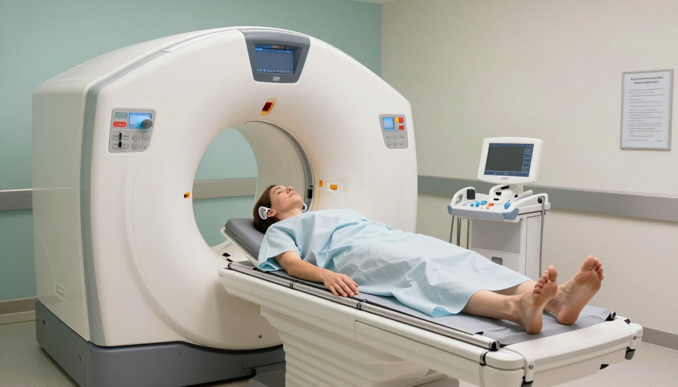
When is a cervical spine MRI recommended?
A cervical spine MRI is often considered when symptoms continue, worsen or suggest a problem involving the nerves or spinal cord. Common reasons include:
- Ongoing neck pain that does not improve with rest, medication or physiotherapy
- Pain travelling from the neck into the shoulder, arm or hand
- Pins and needles, numbness or weakness in an arm or hand
- Suspected slipped disc, nerve compression or spinal stenosis
- Symptoms following an injury or whiplash
- Changes in walking, balance or hand coordination that need investigation
An MRI does not automatically mean that surgery or serious disease is present. It gives a clinician more information about the structures causing symptoms and can support decisions about physiotherapy, pain management, injections, surgery or further tests.
If symptoms are severe, rapidly worsening or accompanied by significant weakness, loss of bladder or bowel control, or difficulty walking, seek urgent medical advice rather than waiting for a private scan.
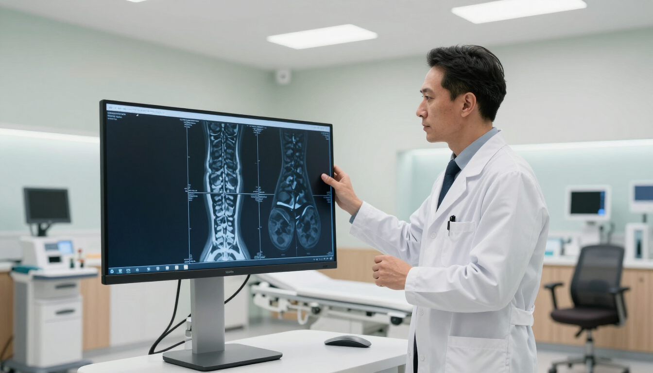
What does a private cervical spine MRI show?
A cervical spine MRI focuses on the neck section of the spine. Depending on the clinical question, images may show:
- The cervical vertebrae and their alignment
- Intervertebral discs and signs of bulging or herniation
- The spinal cord and spaces around it
- Nerve roots and possible areas of compression
- Spinal canal narrowing, also called stenosis
- Inflammation, swelling, infection or other structural changes
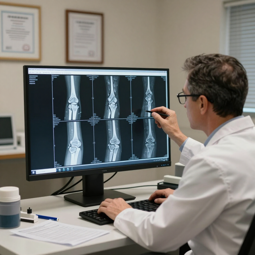
MRI is particularly useful for soft tissues, which may be difficult to assess in detail with an X-ray. A radiologist reviews the images and prepares a written report for the referring clinician.
The scan is one part of an assessment. MRI findings need to be interpreted alongside your symptoms, medical history and physical examination because some changes can occur without causing pain.
How much does a private cervical spine MRI cost in the UK?
The cost of a private cervical spine MRI in the UK commonly falls within the range of several hundred pounds, although the exact price depends on the provider, location, scan type and whether contrast is required. Some clinics publish a fixed self-pay price, while others quote after reviewing a referral.
Before booking, check whether the price includes:
- The MRI scan itself
- A radiologist’s report
- Images supplied digitally or through an online portal
- A copy sent to your GP or consultant
- Contrast, if clinically required
- Any follow-up consultation or administration fee
A lower headline price may not include reporting or a referral review. Ask for the complete cost in writing so you can compare providers fairly. Prices may also differ between London, Leigh-on-Sea and other parts of the UK.
MRI Plus provides diagnostic services designed around accessible booking, clear communication and prompt reporting. Visit the MRI Plus News page for information about the provider and its diagnostic services.
How do you prepare for a private cervical spine MRI?
Most cervical spine MRI scans require little preparation. You can usually eat, drink and take regular medication as normal unless the clinic gives different instructions.
Before your appointment, tell the provider if you:
- Have a pacemaker, cochlear implant, neurostimulator or other implanted device
- Have metal fragments in your body, particularly near your eyes
- Have had surgery involving metal clips, plates or wires
- Are pregnant or may be pregnant
- Have kidney problems or a previous reaction to MRI contrast
- Experience claustrophobia or difficulty lying still
Remove jewellery, watches, piercings and other metal objects before the scan. You may be asked to change into suitable clothing. MRI does not use ionising radiation, but the strong magnetic field means safety screening is essential.
If you are anxious, tell the team in advance. They may explain the process, offer practical reassurance or discuss whether additional support is appropriate.

What happens during a cervical spine MRI scan?
A typical appointment follows these steps:
- Safety check: The radiographer confirms your medical history, implants and any relevant symptoms.
- Changing and positioning: You remove metal items and lie on your back on the scanner table. A support may be placed around your head and neck.
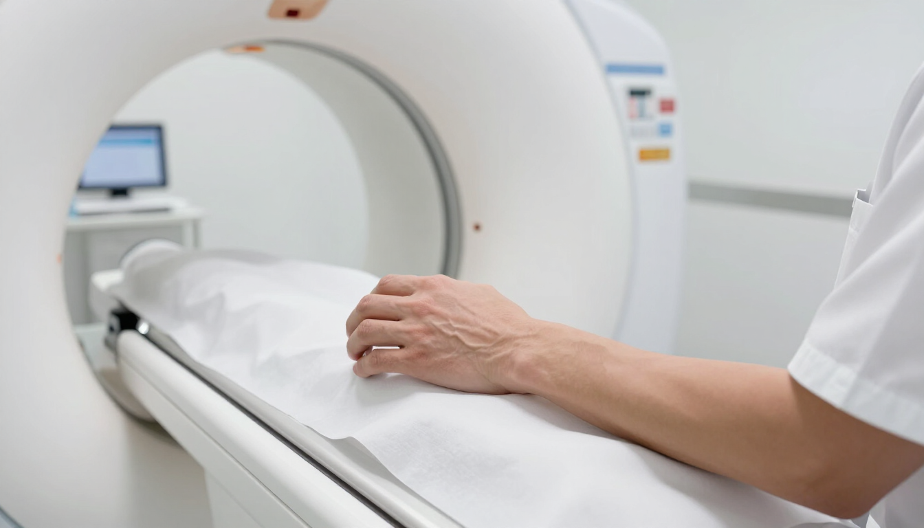
- Image capture: The table moves into the MRI scanner. You will hear loud tapping or knocking sounds, so earplugs or headphones are provided.
- Communication: The radiographer can see and hear you throughout the scan. You may be given a call button.
- Completion: The table moves out, and you can normally leave shortly afterwards.
The scan itself often takes around 20 to 40 minutes, although timings vary. Staying still is important because movement can reduce image quality. You may hear different sequences as the scanner collects images from several angles.
Some examinations use a contrast agent injected into a vein. This is not needed for every neck MRI and depends on the reason for the scan and the radiologist’s or clinician’s request.
How quickly will you receive MRI results?
Private providers may offer faster reporting than standard referral pathways, but turnaround times vary. Ask when the report is expected and how it will be delivered before your appointment.
Results may be sent to your referring GP, consultant or another healthcare professional. Some providers also make images available through a secure digital portal. The report should be discussed with the clinician who understands your symptoms and can explain what the findings mean for your treatment.
A scan report may use terms such as “disc bulge,” “degeneration” or “foraminal narrowing.” These phrases can sound alarming, but their importance depends on their location, severity and relationship to your symptoms. Avoid self-diagnosing from the report alone.
Choosing a private MRI provider
When comparing private cervical spine MRI providers, look beyond price. A suitable service should offer:
- Clear self-pay pricing and an explanation of what is included
- Appropriate clinical referral and safety screening
- Qualified radiography and radiology professionals
- Secure handling of images and personal information
- A realistic reporting timeframe
- Accessible support before and after the appointment
Ask whether the clinic can accommodate your symptoms, mobility needs or anxiety. You should also confirm what happens if the radiologist recommends further assessment. A smooth booking process matters, but clinical governance, accurate reporting and communication are equally important.
For patients in and around London or Leigh-on-Sea, a private diagnostic provider may offer greater flexibility around appointment times. However, the right choice is the provider that combines convenience with safe, properly reported imaging.
Frequently asked questions
Is a cervical spine MRI painful?
No. The scan is painless, although lying still may feel uncomfortable if your neck is already sore. The machine is noisy, but ear protection is provided.
Can I have an MRI if I am claustrophobic?
Often, yes. Tell the clinic when booking. The team can explain the scan, maintain communication and discuss suitable support. Do not take a sedative unless it has been prescribed and arranged safely.
Do I need a referral for a private neck MRI?
Requirements vary. Many providers need a referral or clinical request so the correct scan can be performed and reported safely. Ask the clinic what information it requires before booking.
Is MRI better than an X-ray for neck pain?
They answer different questions. X-rays show bones and alignment, while MRI provides much more detail about discs, nerves, the spinal cord and other soft tissues. A clinician can advise which test is appropriate.
Should I choose contrast for my scan?
Not usually without clinical advice. Contrast is used for specific questions and is not required for every cervical spine MRI. The referring clinician or radiologist will decide whether it is appropriate.
Key takeaways
A private cervical spine MRI UK scan can be useful when persistent neck pain, radiating arm pain, numbness, weakness or other neurological symptoms do not improve with basic treatment. It provides detailed images of discs, nerves and the spinal cord, helping a clinician plan the next step. Costs vary, so confirm whether reporting, images and contrast are included. Prepare by completing the safety screening and disclosing implants or medical conditions. Compare providers on clinical quality, communication and turnaround time—not price alone. If symptoms are severe or rapidly worsening, seek urgent medical advice rather than waiting for a routine appointment.