What Is an MRCP Scan?
An MRCP scan is a non-invasive MRI examination that creates detailed images of the bile ducts, pancreatic duct, gallbladder and surrounding organs. Doctors use this type of MRI scan to investigate pain, jaundice, gallstones, inflammation, narrowing and other conditions without using an endoscope or X-rays.
What is an MRCP scan?
MRCP stands for magnetic resonance cholangiopancreatography . It is a specialised form of MRI that focuses on the body’s biliary and pancreatic systems.
Unlike some investigations, an MRCP scan does not require a camera to be passed into the digestive tract. Instead, the MRI scanner uses a strong magnetic field and radio waves to produce detailed images. These can help clinicians assess:
- The liver and gallbladder
- The bile ducts
- The pancreatic duct
- The pancreas
- Surrounding abdominal structures
An MRCP is usually an outpatient examination. This means you can generally return home after the scan unless your healthcare team advises otherwise.
Why is an MRCP scan used?
An MRCP MRI can give doctors a clearer view of structures that may be difficult to assess with other imaging tests. It is often requested when symptoms, blood tests or previous scans suggest a problem involving the bile ducts, gallbladder or pancreas.
Common reasons for an MRCP include investigating:
- Gallstones or stones in the bile duct
- Bile duct blockage or narrowing
- Unexplained jaundice
- Recurrent abdominal pain
- Pancreatitis or suspected inflammation
- Cysts, tumours or other structural changes
- Abnormal liver or pancreatic blood-test results
The scan may also help a consultant decide whether further treatment or an investigation such as endoscopy is needed. It does not replace every other test, but it can provide valuable information without the need for an invasive procedure.
How does an MRCP compare with other tests?
Your clinician will choose the most appropriate test based on your symptoms and medical history. An MRCP scan is different from several commonly used investigations:
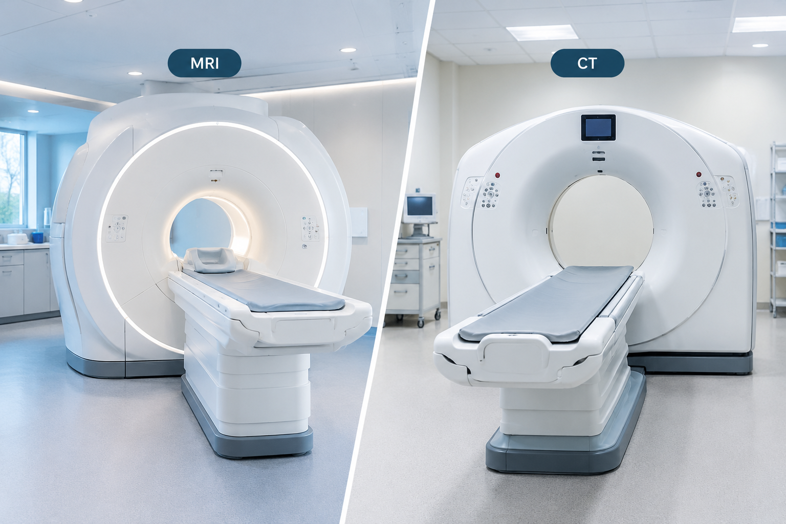
- MRI: A specialised type of MRI scan that uses a magnetic field and radio waves, with no radiation. It gives detailed images of the bile and pancreatic ducts without needing instruments to be passed into the body, making it a non-invasive option for assessing the ducts.
- Ultrasound: Uses sound waves and is often useful for assessing the gallbladder, but it may not show every duct clearly.
- CT scan: Uses X-rays and can provide detailed cross-sectional images, although it may offer less specialised visualisation of the bile and pancreatic ducts.
- Endoscopic retrograde cholangiopancreatography (ERCP): Uses an endoscope and can treat some blockages, but it is more invasive and carries procedure-related risks.
- Gastroscopy or colonoscopy: Examines the digestive tract from inside and is not designed to provide the same view of the ducts.
An MRCP is primarily a diagnostic scan. If treatment is required, your consultant will explain the next steps.
How should you prepare for an MRCP MRI?
Preparation instructions can vary between clinics, so always follow the guidance provided with your appointment. In many cases, you will be asked not to eat for several hours before the scan. This can help reduce movement from digestion and improve the quality of the images.
Before attending, you should:
- Check whether you need to fast and confirm when you may last eat or drink.
- Take regular medication only as advised by your healthcare team.
- Tell the clinic if you are pregnant, might be pregnant or are breastfeeding.
- Tell staff about kidney problems, allergies, previous surgery or any implanted medical devices.
- Wear comfortable clothing without metal fasteners where possible.
You may be asked to complete a safety questionnaire. MRI uses a powerful magnet, so it is essential to disclose any pacemaker, cochlear implant, metal fragment or other device. Some implants are MRI-compatible, but the team must check this before scanning.
For further information about available diagnostic services, visit the MRI Plus.
What happens during the MRCP scan procedure?
The exact process may vary slightly, but an MRCP scan commonly follows these steps:
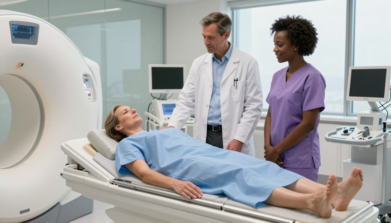
- Safety checks: A radiographer confirms your details, medical history and MRI safety information.
- Changing: You may need to remove jewellery, watches, hearing aids, cards and clothing containing metal.
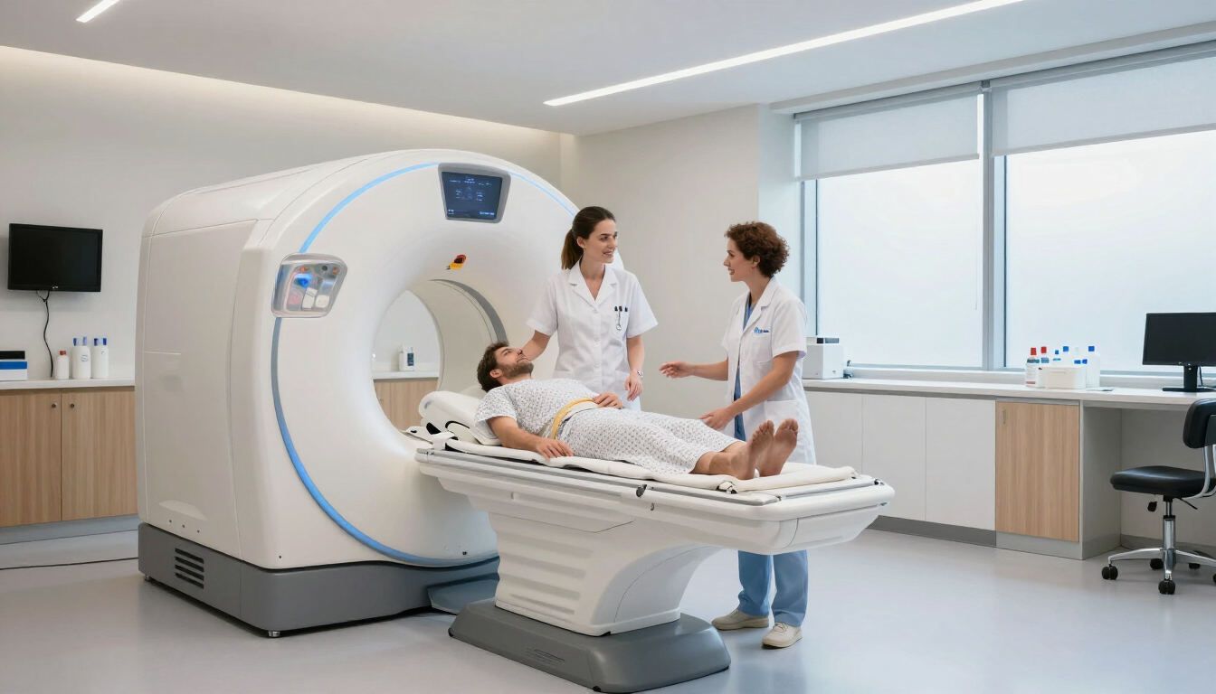
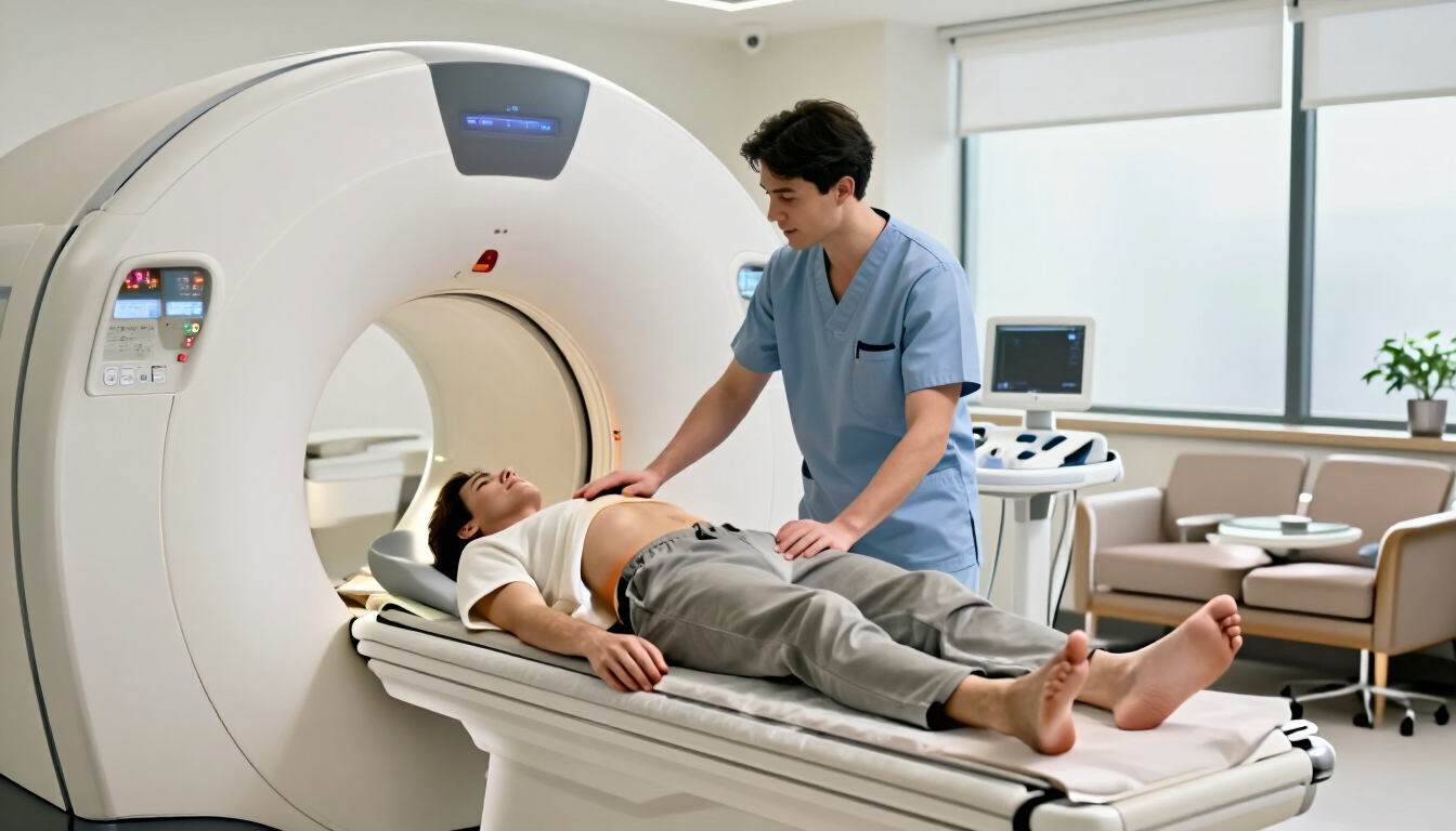
- Positioning: You lie on your back on a padded scanner table. A device may be placed over your abdomen to help create clear images.
- Scanning: The table moves into the MRI scanner. You will need to remain still while a series of images is taken.
- Breath-holds: The radiographer may ask you to hold your breath briefly at certain points.
- Completion: Once the images are checked, you can usually leave and continue your day.
The scanner makes loud knocking or tapping sounds. You may receive headphones or ear protection, and you can communicate with the radiographer throughout the examination. If you feel uncomfortable, tell the team immediately.
How long does an MRCP take?
The scan itself often takes around 20 to 45 minutes, although the total appointment may be longer because of registration, changing, safety checks and positioning. Timing can depend on the number of images required and whether contrast is recommended.
Most people find the scan straightforward, although staying still in a confined space can feel challenging. If you have concerns about claustrophobia, discuss them with the clinic before your appointment. The team may be able to provide reassurance or explain available support.
Is contrast used for an MRCP scan?
Many MRCP examinations are performed without contrast because fluid in the bile and pancreatic ducts can appear clearly on specialised MRI sequences. In some situations, your clinician or radiologist may recommend contrast to provide additional information.
If contrast is being considered, staff will ask about kidney function, allergies and previous reactions. Do not assume contrast is required or unsuitable: your clinical team will make an individual decision based on the reason for the scan.
Are there risks or side effects?
An MRCP scan does not use ionising radiation. MRI is generally considered safe when appropriate screening has been completed. The main considerations are related to metal devices, implanted materials, pregnancy and, where used, contrast medication.
Some people experience temporary discomfort from lying still, noise or the enclosed scanner. Serious reactions are uncommon, but you should report any unusual symptoms to the imaging team promptly.
When will you receive MRCP results?
A radiologist reviews the images and prepares a report for the doctor or consultant who requested the scan. The timescale depends on the clinic and the urgency of the referral. Your healthcare professional will explain the findings and recommend any follow-up.
The radiographer performing the examination may not be able to interpret or discuss the results immediately. If you have questions, ask when and how your report will be shared.
Frequently asked questions
Can I drive after an MRCP scan?
Usually, yes. If you have not received sedation or medication that affects alertness, you can generally drive and return to normal activities. Follow the instructions given by your clinic.
Is an MRCP painful?
The scan is not usually painful. You must lie still, and the scanner is noisy, but the examination is non-invasive. Tell the radiographer if you feel discomfort.
Can an MRCP detect cancer?
An MRCP can show abnormalities such as masses, narrowing or changes in the bile and pancreatic ducts. It cannot always provide a definitive diagnosis. Further tests may be needed.
Can I eat before an MRCP scan?
You may need to avoid food for several hours beforehand. Instructions differ, so follow the specific preparation guidance from your imaging provider.
Key takeaways
An MRCP scan is a non-invasive MRI examination used to assess the bile ducts, pancreatic duct, gallbladder, pancreas and nearby structures. It can help investigate symptoms such as abdominal pain, jaundice, suspected gallstones and pancreatitis.
Preparation commonly involves fasting and completing MRI safety checks. During the procedure, you lie still in the scanner while images are taken, sometimes with brief breath-holds. The examination is usually comfortable and does not use X-ray radiation.
If your clinician has recommended an MRCP MRI, check your appointment instructions carefully, disclose implants or medical conditions, and ask when your results will be discussed.