Foot and Ankle MRI Guide: 5 Ways to Diagnose Foot Pain
A foot and ankle MRI is a non-invasive test. It uses magnetic fields and radio waves to create detailed images. These images show the complex structures in the lower limb. It is the gold standard for finding soft tissue injuries and stress fractures. It also helps detect inflammatory conditions that often do not show on X-rays.
Why is foot pain diagnosis so critical?
Foot pain is more than just a minor inconvenience; it is a fundamental disruption to a patient’s mobility and overall quality of life. For podiatrists, physiotherapists or sports medicine specialists, the first step in a successful rehabilitation plan is to identify the causes of the foot pain. The human foot is a marvel of engineering, comprising 26 bones, 33 joints, and over a hundred muscles, tendons, and ligaments. Because these structures are so densely packed, pain in one area often radiates from another, making a clinical physical exam sometimes inconclusive.
When a patient presents with chronic pain, it often indicates an underlying mechanical failure or inflammatory process. Ignoring these signals can lead to compensatory movements, where the patient alters their gait to avoid pain. This shift frequently results in secondary injuries in the knee, hip, or lower back. A full-colour Foot and Ankle MRI allows practitioners to differentiate a simple strain from something more sinister, such as bone marrow oedema or a high-grade ligament tear. Proper imaging allows for early intervention to prevent long-term disability and to get the patient back to daily and athletic activities as safely as possible.
“In the UK healthcare landscape, many patients are waiting too long for these critical diagnostics.” MRI Plus News discusses how rapid access to imaging can shift the pathway of patient care from a "wait and see" mentality to proactive management. Early diagnosis relieves the patient's psychological distress, and gives the patient a clear answer and a way to recovery.
Anatomy of the Foot and Ankle
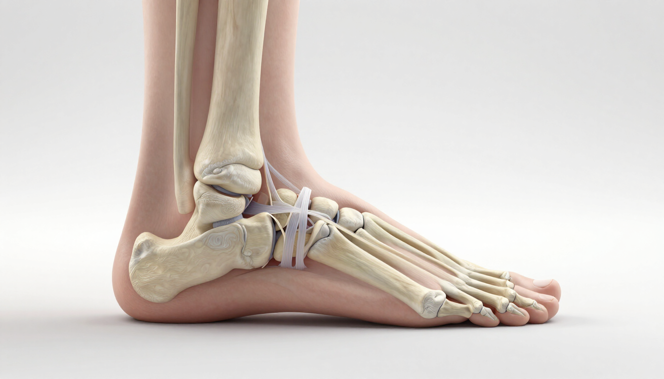
To appreciate the diagnostic power of an MRI, one must appreciate the complex landscape it visualises. The foot is traditionally divided into three main parts, the forefoot, midfoot and hindfoot. MRI captures with incredible clarity the specific anatomical landmark of each area.
- The Hindfoot: This includes the talus (ankle bone) and the calcaneus (heel bone). The MRI is essential here for assessing the subtalar joint and the crucial Achilles tendon, the strongest tendon in the body. Injuries here, such as Haglund’s deformity or retrocalcaneal bursitis, are common causes of posterior heel pain.
- The Midfoot: A complex cluster of small bones—the navicular, cuboid, and three cuneiforms. This area is the site of the Lisfranc ligament complex. A Lisfranc injury is notoriously difficult to diagnose with X-ray alone but is clearly visible on a high-resolution MRI.
- The Forefoot: Comprising the metatarsals and phalanges (toes). This is a frequent site for stress fractures and Morton’s neuroma, a painful thickening of the tissue around the nerves leading to the toes.
- Ligamentous Structures: The ankle is supported by various ligament groups, most notably the lateral collateral ligaments (including the ATFL and CFL) and the medial deltoid ligament. MRI is the only modality that can accurately grade the severity of tears in these structures.
- Tendon Groups: Beyond the Achilles, the peroneal tendons on the lateral side and the posterior tibial tendon on the medial side are critical for foot stability. MRI helps identify tenosynovitis or longitudinal split tears that can lead to adult-acquired flatfoot deformity.
Top benefits of a foot and ankle MRI scan
The transition from traditional imaging to MRI offers several transformative benefits for both the clinician and the patient. While X-rays remain useful for checking basic bone alignment or obvious fractures, they fall short in many areas where MRI excels.
- Superior Soft Tissue Contrast: MRI gives clear detail of soft tissues. It can tell a sprain from a partial tear. It can also spot a complete rupture. This helps decide between rest and surgery.
- Early Detection of Stress Reactions: Before a stress fracture shows on an X-ray, the bone is stressed. This can cause marrow swelling. MRI can spot these changes weeks earlier. This lets athletes rest before a major injury.
- No Ionising Radiation: Unlike CT scans or X-rays, MRI uses magnets and radio waves. It has no ionising radiation. This makes it safer for repeat scans. It is also better for younger patients. It suits people with long-term conditions.
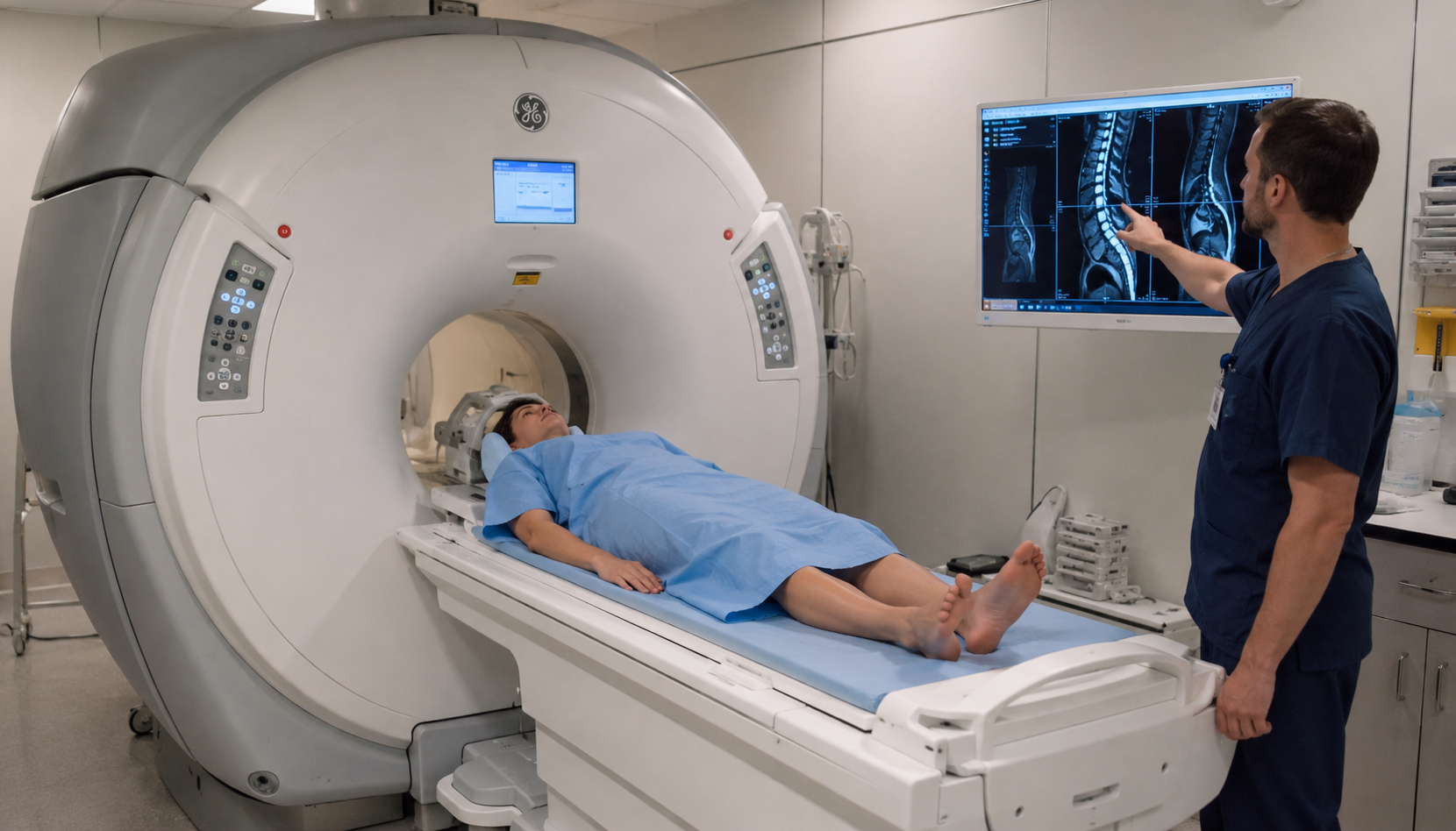
- Comprehensive 3D Visualisation: MRI takes images in several planes. These include axial, sagittal, and coronal. Doctors can view the foot from many angles. This helps avoid missing small lesions behind bones.
- Detection of Occult Pathologies: Foot pain can come from ganglion cysts or hidden tumours. It can also be from early infection, like osteomyelitis. MRI is very sensitive to these issues. It can reassure both patient and clinician.
When should you recommend a foot and ankle MRI?
Determining the right time to order an MRI is a clinical art. Generally, if a patient has undergone 2-4 weeks of conservative treatment (rest, ice, physical therapy) without significant improvement, advanced imaging is warranted. This is particularly true if the pain is localized to a specific joint or if there is persistent swelling without a clear cause.
For sports-related injuries, the timeline is often compressed. An athlete who cannot bear weight or has significant instability after an inversion injury should be considered for an MRI sooner rather than later to rule out syndesmotic (high ankle) sprains. Additionally, patients with diabetes who present with foot pain should be monitored closely; MRI is the preferred method for diagnosing Charcot foot in its early stages, where prompt treatment can prevent limb-threatening complications. Professionals who follow a Foot and Ankle MRI Guide recognize that imaging is not just about finding the "what," but also understanding the "where" and "how much" regarding tissue damage.
How does an MRI compare to other imaging?
It is common for patients to ask why they need an MRI if they have already had an X-ray or an Ultrasound. Each tool has its place in the diagnostic hierarchy, but they serve different purposes. X-rays are fantastic for "hard" structures—bones and joints—and are usually the first line of defense due to their low cost and speed. However, an X-ray is essentially a 2D shadow; it cannot see the inflammation within a bone or the fraying of a tendon.
Ultrasound is another excellent tool, particularly for dynamic imaging (watching a tendon move in real-time). It is cost-effective and does not require the patient to sit in a tunnel. However, ultrasound is highly operator-dependent and cannot penetrate bone to see what is happening inside a joint or deep within the midfoot bones. This is where the MRI becomes indispensable. It bridges the gap by providing the high-resolution internal view that ultrasound lacks and the soft-tissue depth that X-ray cannot provide. For a clinician, the MRI is the definitive "final word" in the diagnostic process.
Clinical Insights for Healthcare Professionals
When reviewing an MRI report, it is important to correlate the findings with the patient's clinical symptoms. An MRI might show a small labral tear or a minor tendon thickening that is actually asymptomatic and unrelated to the patient's primary complaint. This is why a thorough physical examination remains the foundation of diagnosis. The MRI should be viewed as a tool to confirm or refine a clinical hypothesis, not as a replacement for clinical judgment.
At MRI Plus, the focus is on providing these insights rapidly. We understand that in the world of professional sports and busy clinical practices, time is the most valuable resource. By offering rapid appointment availability and prompt reporting, we ensure that the diagnostic loop is closed quickly. This efficiency allows the physiotherapist or consultant to begin the correct treatment path immediately, whether that involves custom orthotics, targeted injections, or specialized exercise protocols.
How can patients prepare for a foot MRI scan?
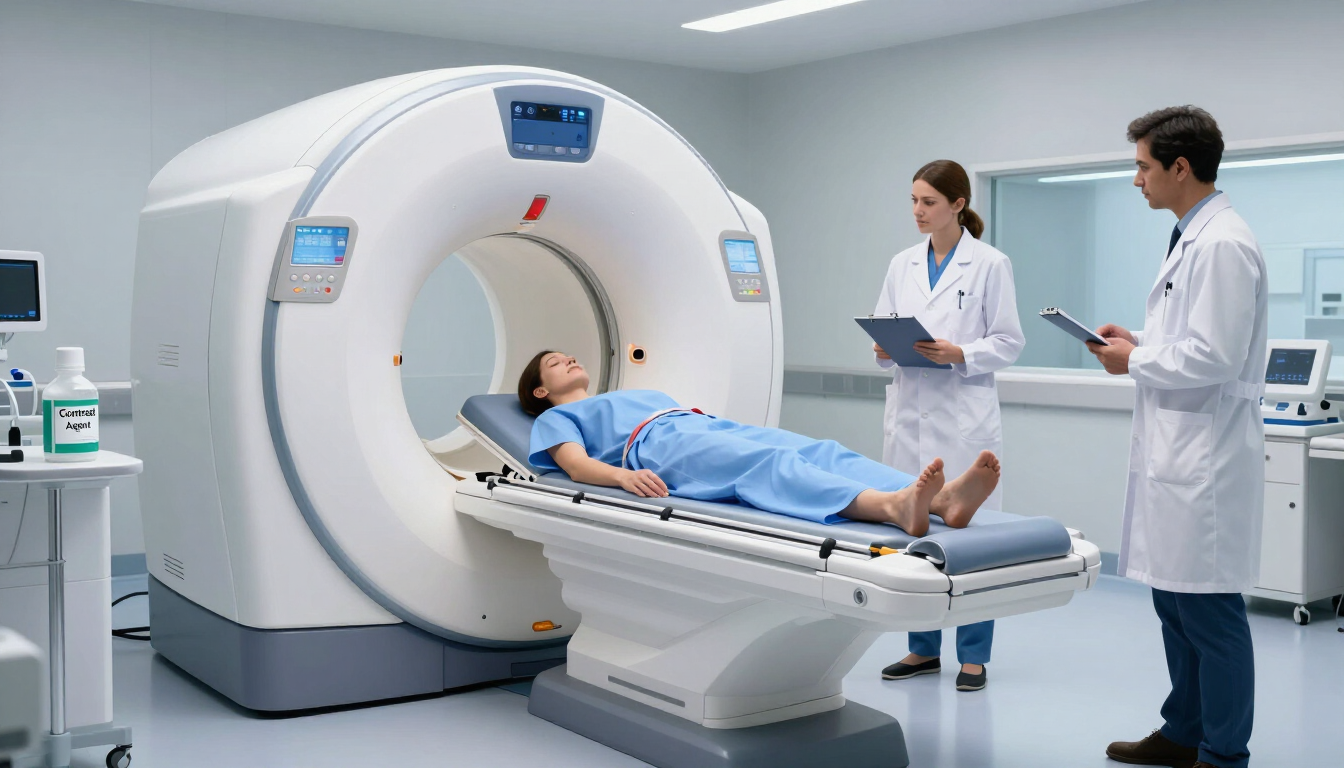
Preparation for a foot and ankle MRI is quite simple. Clear communication can help reduce patient anxiety. Patients should wear loose, comfortable clothing. They should avoid jewellery or metal accessories. The MRI’s strong magnetic field can react with metal. If the patient has any implants, they must tell the facility in advance. Examples include pacemakers, cochlear implants, and some surgical clips.
During the scan, the patient will lie on their back. The affected foot will be placed in a special “coil”. This acts like a camera to capture the images. The scan usually takes 20 to 45 minutes. Since the foot is far from the head, many claustrophobic patients find this easier. It is often easier than head or chest scans. Sharing this information early can improve the patient’s experience. It can also help the technician get clear images without motion blur.
Summary and Takeaways
A foot and ankle MRI guide serves as an essential resource for professionals looking to optimise patient outcomes. By providing unmatched detail of soft tissues, early bone stress detection, and a clear view of complex joint mechanics, the MRI removes the guesswork from foot pain diagnosis. Whether you are managing a weekend warrior with a suspected Achilles tear or a professional athlete with a midfoot injury, rapid access to high-quality imaging is the key to a fast recovery.
- MRI is the gold standard for soft tissue and occult bone injury detection.
- Timing matters: Move to MRI if conservative therapy fails after 2-4 weeks.
- Clinical correlation is vital; treat the patient, not just the image.
- Efficiency wins: Fast diagnostics lead to faster rehabilitation and better patient satisfaction.
In our experience treating hundreds of orthopedic referrals, foot and ankle pain is one of the most frequently misdiagnosed conditions in primary care — often because plain X-rays miss soft tissue, ligament, and cartilage damage. MRI closes that diagnostic gap, giving your patients (and you) a clear, evidence-based picture before deciding on treatment.
At MRI Plus, our board-certified radiologists and accredited imaging centres are built specifically to support referring physicians with fast turnaround and clear, actionable reports not just images.
References :
BOFAS > Hyperbook > Radiology > 9. Advanced Imaging. (n.d.). https://www.bofas.org.uk/hyperbook/radiology/9-advanced-imaging
Preview. (n.d.). https://acsearch.acr.org/docs/69424/Narrative/
Boa. (n.d.). Commonly missed injuries in the foot and ankle. https://www.boa.ac.uk/resource/commonly-missed-injuries-in-the-foot-and-ankle.html