Wrist Ligament Tear MRI: Symptoms, Diagnosis & What the Scan Shows
A wrist ligament tear MRI is the primary diagnostic tool used to identify soft tissue damage within the complex wrist joint. By utilizing high-frequency magnetic fields, it provides detailed visualization of ligaments, tendons, and cartilage, effectively detecting injuries like TFCC or scapholunate tears that standard X-rays frequently overlook.
What are the symptoms of a wrist ligament tear?
Identifying a ligament tear in the wrist early is crucial for preventing long-term joint instability and degenerative conditions like arthritis. Because the wrist is composed of eight small carpal bones held together by an intricate network of ligaments, even a small tear can significantly disrupt hand function. Patients often describe a range of symptoms that vary depending on which specific ligament is affected, but several universal warning signs suggest a specialized scan is necessary.
One of the most common symptoms is persistent pain that does not improve with rest. While a simple strain might settle within a few days, a ligament tear often results in a deep, aching sensation that intensifies when you try to grip objects or twist your wrist, such as when opening a jar or turning a door handle. This pain is often localised; for instance, pain on the thumb side of the wrist often indicates a scapholunate injury, while pain on the pinky side (the ulnar side) suggests a TFCC issue.
Beyond pain, you might notice physical changes in the joint’s behavior. Many patients report a "clicking," "popping," or "clunking" sensation during movement. This occurs because the bones are no longer being held in their proper alignment, causing them to shift abnormally against one another. You should also watch for:
- Significant swelling and bruising shortly after an injury.
- A noticeable loss of grip strength or hand weakness.
- A feeling of instability, as if the wrist might "give way."
- Reduced range of motion, particularly when bending the wrist back.
- Tenderness when pressing directly on the joint line.
If you are experiencing these issues, keeping up with MRI Plus News can help you understand the latest advancements in diagnostic imaging and how rapid access to scans can change your recovery trajectory.
Common types of wrist ligament injuries
The wrist is a marvel of biological engineering, but its complexity makes it vulnerable to several specific types of ligamentous trauma. Understanding which structure is injured is the first step toward a successful recovery. We frequently see patients in London and Leigh-on-Sea who are suffering from one of the following conditions, all of which require precise imaging for a formal diagnosis.
The Scapholunate Ligament Tear is arguably the most common and clinically significant. This ligament connects the scaphoid and lunate bones. When it tears, these bones begin to move apart, leading to a condition known as DISI (Dorsal Intercalated Segment Instability). Without an accurate diagnosis via MRI, this can lead to progressive wrist collapse over time. Another frequent injury involves the Triangular Fibrocartilage Complex (TFCC). The TFCC acts as a stabiliser for the ulnar side of the wrist and provides a cushion for the carpal bones. Tears here are common in athletes who perform repetitive twisting motions, such as tennis players or golfers.
Other less common but equally debilitating injuries include Lunotriquetral ligament tears and injuries to the extrinsic ligaments that connect the forearm bones to the hand. Each of these injuries presents differently and requires a tailored approach to rehabilitation. A high-quality scan ensures that your consultant isn't guessing but is instead working with a clear map of your internal anatomy.
Why is an MRI better than an X-ray for wrist pain?
When you first visit a doctor for wrist pain, they will often request an X-ray. While X-rays are excellent for detecting fractures or bone dislocations, they have significant limitations when it comes to the wrist's soft tissues. Ligaments, tendons, and cartilage are essentially "invisible" on a standard X-ray because they do not have the density required to block the radiation. An X-ray might show that your bones are in the right place, but it cannot show the status of the "cables" holding them together.
A wrist ligament tear MRI, however, uses magnetic resonance to excite hydrogen atoms in your body's water molecules. Because soft tissues like ligaments have a high water content compared to bone, the MRI can create a high-contrast image that clearly differentiates between healthy tissue and a tear. This is particularly important for "occult" injuries that are present but hidden from traditional diagnostic tools.
Furthermore, MRI technology allows for multi-planar imaging. This means the radiologist can look at your wrist from the front, the side, and even in cross-sections. This three-dimensional perspective is vital for determining the severity of a tear—whether it is a partial fraying of the ligament or a complete rupture. In many cases, an MRI will also reveal "bone bruising" (bone marrow oedema), which provides additional clues about the mechanism of the injury and the stress the joint has undergone.
What does a wrist ligament tear look like on an MRI?
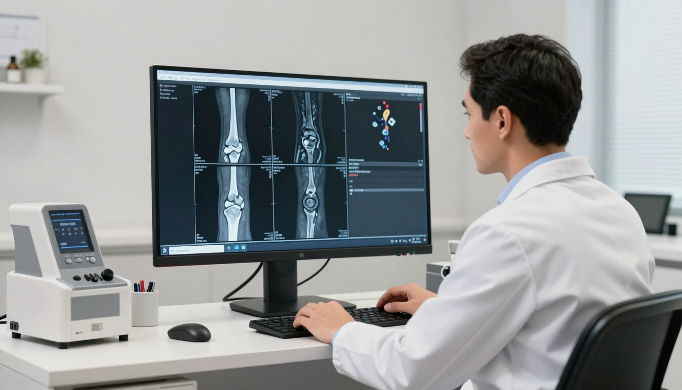
For a trained radiologist, a wrist ligament tear MRI reveals specific visual markers that indicate trauma. In a healthy wrist, ligaments appear as dark, tight bands of tissue connecting the white or grey bones. When a tear occurs, this dark band may appear disrupted, wavy, or entirely absent. One of the primary indicators of a recent tear is the presence of "high signal intensity." In MRI terminology, this means an area appears bright white on certain types of scans, usually indicating fluid or inflammation that has rushed to the site of the injury.
In the case of a scapholunate tear, the scan may show a widened gap between the scaphoid and lunate bones, often filled with joint fluid. For a TFCC tear, the radiologist looks for irregularities in the disc-shaped structure on the pinky side of the wrist. Sometimes, a "contrast agent" (gadolinium) may be injected into the joint—a procedure known as an MR Arthrogram—to make even the smallest leaks or tears even more visible. This level of detail is why private diagnostics are so highly valued; they provide the clarity needed for a definitive surgical or non-surgical plan.
The MRI scanning process at MRI Plus
At MRI Plus, we understand that medical imaging can be an intimidating prospect for many patients. That is why our process is designed around speed, transparency, and patient comfort. When you arrive at our clinic, you will be greeted by our clinical team who will guide you through a safety screening to ensure you have no metal implants that could interfere with the magnetic field. Unlike older, cramped machines, modern MRI units are designed to be more spacious and less claustrophobic.
For a wrist scan, you will usually lie on your back or side, and your arm will be positioned inside a specialized "wrist coil." This coil acts like a high-powered antenna, focusing the magnetic field specifically on the small area of your wrist to produce the sharpest possible images. The scan itself typically takes between 30 and 45 minutes. During this time, you will hear a series of loud rhythmic noises—this is completely normal and is simply the machine’s magnets switching on and off. We provide headphones or earplugs to make the experience as quiet as possible.
The efficiency of our service means you don't have to wait weeks for an appointment. We prioritize rapid access, knowing that every day spent waiting for a diagnosis is a day lost in your recovery. Our patients in Leigh-on-Sea and London appreciate that they can book online and receive professional care without the friction often found in traditional healthcare pathways.
How long does it take to get MRI results?
One of the biggest frustrations in healthcare is the "waiting game" for results. At MRI Plus, we leverage modern digital tools to ensure that your images are reviewed by a specialist consultant radiologist as quickly as possible. In most cases, a detailed clinical report is delivered to you and your referring clinician within 24 to 48 hours. This report will detail the findings, confirm the presence and location of any tears, and offer a clinical opinion on the state of the joint.
Having these results quickly allows you to move straight to the next phase of your care. Whether that involves a consultation with an orthopaedic surgeon or starting a specialized hand therapy programme, you can move forward with the confidence that you are treating the correct issue. Speed in reporting is central to our clinical governance, ensuring that patient outcomes are optimized through timely intervention.
Next steps after your diagnosis
Once your wrist ligament tear MRI has confirmed the nature of your injury, the next steps depend on the severity of the findings. Not every tear requires surgery. Minor or partial tears often respond well to conservative treatments, including:
- Splinting or casting to immobilize the joint and allow the ligament to heal.
- Specialized hand physiotherapy to strengthen the surrounding muscles.
- Activity modification to avoid aggravating the injury.
- Anti-inflammatory medication to manage pain and swelling.
- Follow-up scans to monitor the healing process.
For complete ruptures or cases where the wrist bones have become unstable, a surgical consultation may be necessary. Modern minimally invasive techniques, such as wrist arthroscopy, allow surgeons to repair ligaments with very small incisions. Regardless of the path, the information provided by your MRI is the foundation upon which your entire recovery is built.
Summary of Wrist Ligament Recovery
A wrist ligament tear can be a debilitating injury, but a dedicated Wrist MRI scan offers the quickest path to clarity and recovery. By identifying the exact location and severity of a tear, patients can avoid the pitfalls of misdiagnosis and long-term joint damage. We provide a streamlined, patient-centric alternative for those seeking fast and accurate diagnostics in the UK.
Key Takeaways:
- MRI is the only definitive way to visualize soft tissue tears in the wrist joint.
- Symptoms like clicking, instability, and localized pain should not be ignored.
- Fast diagnostic reporting (within 48 hours) enables quicker treatment and better outcomes.
- A clear diagnosis allows for a targeted recovery plan, whether conservative or surgical.
If you are struggling with persistent wrist pain, consider booking a private scan to get the answers you need today..
FAQ's :
How long does it take for a torn wrist ligament to heal?
It lists "suspected ligament or tendon injury" as one reason someone might be referred for a wrist MRI, and notes that ligaments and tendons are something the scan can assess, but there's no section covering how long these injuries take to heal. A Wrist ligament tears are usually graded by severity, and healing time follows the grade: Grade 1 (stretched, not torn): around 1 to 3 weeks Grade 2 (partial tear): roughly 3 to 6 weeks Grade 3 (complete tear): the ligament itself usually heals in around 8 to 12 weeks.
Which is better for diagnosing ligament damage, ultrasound or MRI?
MRI is generally best sharper soft-tissue detail, sees deep or intra-articular ligament tears. Ultrasound suits superficial ligaments and real-time movement, and is cheaper/faster, but more operator-dependent.
Can a torn wrist ligament heal by itself?
yes, for mild to moderate tears. With rest, splinting and time, immobilizing a partial tear with a cast or splint can help it heal on its own, and stable injuries usually heal well this way without surgery.