Can an MRI Detect Parasites in the Brain?

Rahul Panchal • July 1, 2026

Yes, an MRI is the gold standard for detecting parasites in the brain. It provides high-contrast images that reveal cysts, larvae, and the specific stage of infection. While CT scans detect calcification, MRI is far superior for identifying active, soft-tissue parasitic lesions and the surrounding inflammatory response.

Can an MRI detect parasites in the brain?

If you are experiencing unexplained neurological symptoms like seizures, severe headaches, or sudden confusion, you might wonder if a hidden infection is to blame. The answer is a definitive yes: a Magnetic Resonance Imaging (MRI) scan is the most effective tool for finding parasites in the brain.

Parasites often enter the body through contaminated food or water, eventually traveling through the bloodstream to find a host. The brain is a particularly sensitive environment where these "uninvited guests" can cause significant damage if not caught early. An MRI for brain parasites works by using powerful magnets and radio waves to create detailed slices of brain tissue. Unlike other imaging methods, the MRI can distinguish between the parasite itself, the protective cyst it might live in, and the fluid or swelling (oedema) that the brain produces in response to the invader.

Radiologist analyzing a detailed brain scan

Why MRI is the Best for Neurological Parasites

When doctors suspect a brain infection, they need more than just a blurry picture; they need a precise map. This is where MRI outperforms other diagnostic tools like CT scans. While a CT scan is excellent for spotting old, calcified (dead) parasites, it often misses active ones that are still causing symptoms.

MRI is considered the best diagnostic method for several reasons:

  • Superior Soft Tissue Contrast: MRI can clearly show the difference between healthy brain matter and a parasitic cyst.
  • Identifying the 'Scolex': In many cases, an MRI can actually show the head of the parasite (the scolex) inside a cyst, which is a “smoking gun” for diagnosis.
  • Visualising Inflammation: It highlights the exact area where the body’s immune system is attacking the parasite, helping doctors understand how much pressure is on the brain.
  • Multiple Sequences: Radiologists use different

Reference :  

World Health Organization. (2021). EVIDENCE PROFILES. WHO Guidelines on Management of Taenia Solium Neurocysticercosis - NCBI Bookshelf. https://www.ncbi.nlm.nih.gov/books/NBK573857/


UCLH / Hospital for Tropical Diseases – UK Parasitology Service : https://www.uclh.nhs.uk/our-services/find-service/pathology-1/parasitology


Brain and Head MRI Scans from £199 | MRI Plus. (n.d.). https://mriplus.co.uk/body-parts/brain-and-head

Foot and Ankle MRI
By Rahul Panchal July 2, 2026
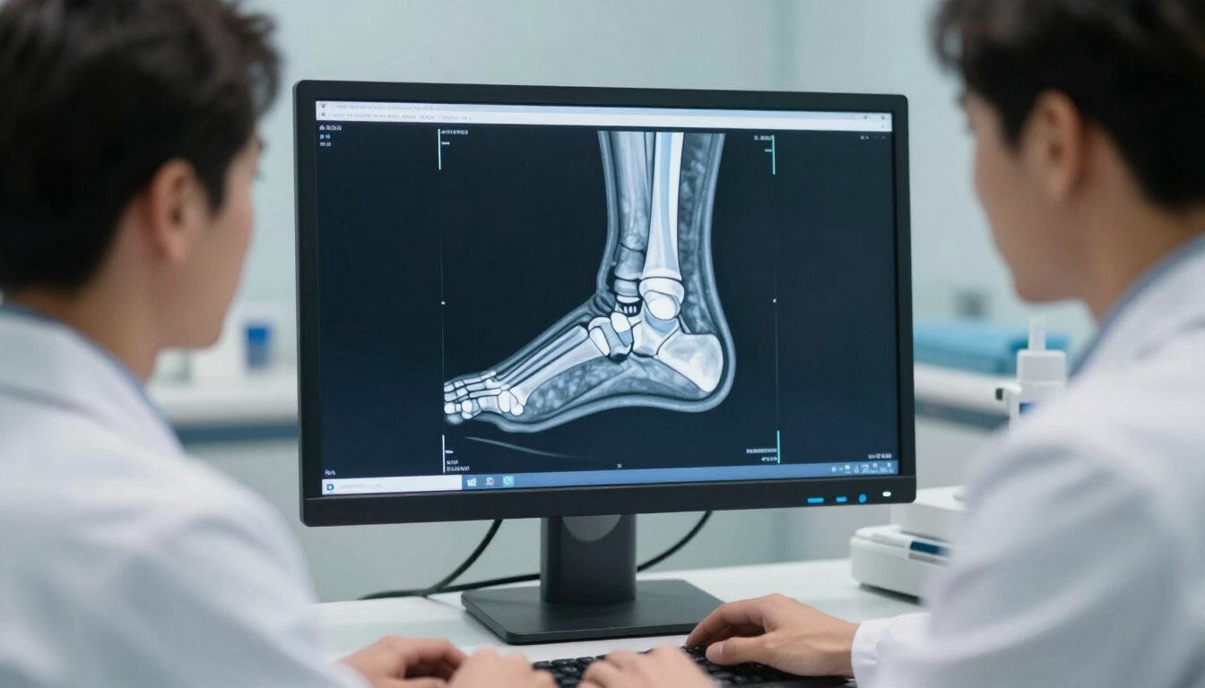
Discover the ultimate foot and ankle MRI guide for professionals. Learn about anatomy, benefits, and how to get rapid diagnostic results for patient foot pain.
Internal Auditory Meatus MRI scan machine in a professional medical setting.
By Rahul Panchal June 30, 2026
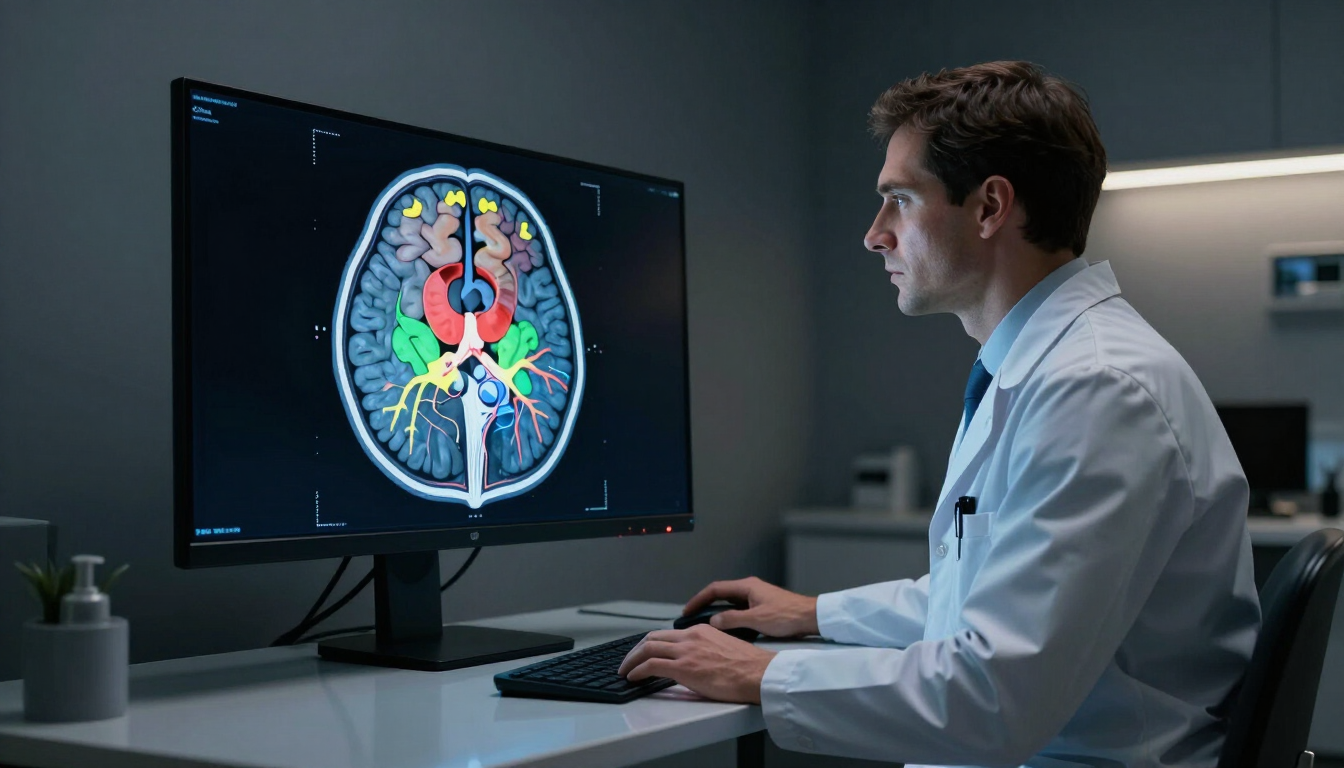
Learn what an Internal Auditory Meatus (IAM) MRI scan is, when it is needed for hearing loss or tinnitus, and how to book a private ear MRI scan today.
MRI With Contrast
By Rahul Panchal June 29, 2026
Preparing for an MRI with contrast? Learn what happens before, during, and after your scan, including contrast dye information, preparation tips, and what to expect.
Full Spine MRI Scan
By Rahul Panchal June 26, 2026
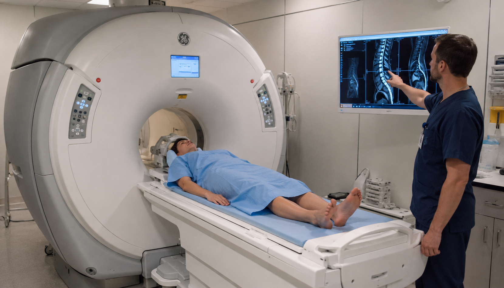
Discover what a full spine MRI scan shows, the duration, and private costs in the UK. Book a rapid spine MRI with MRI Plus for fast, professional results.