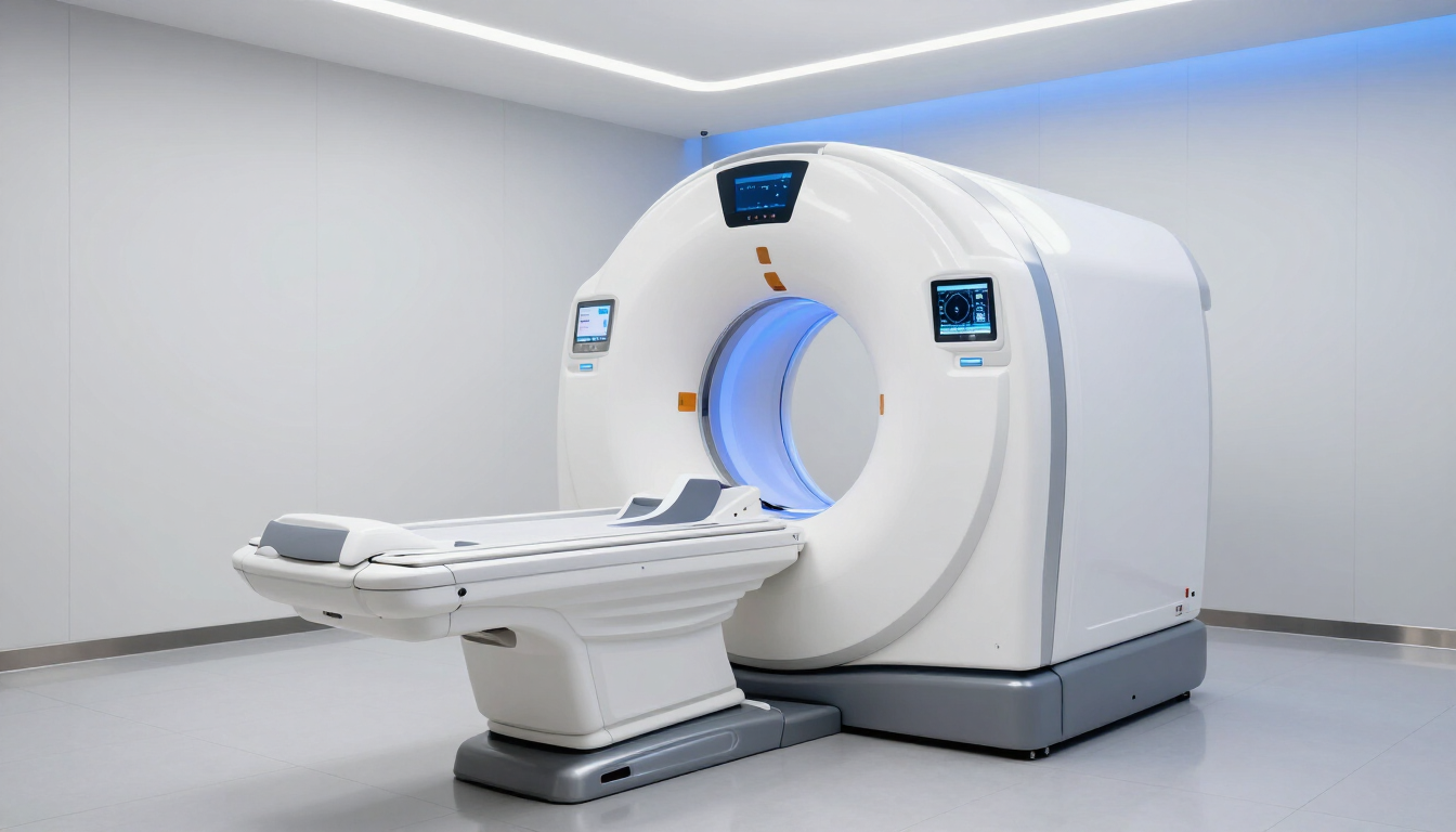
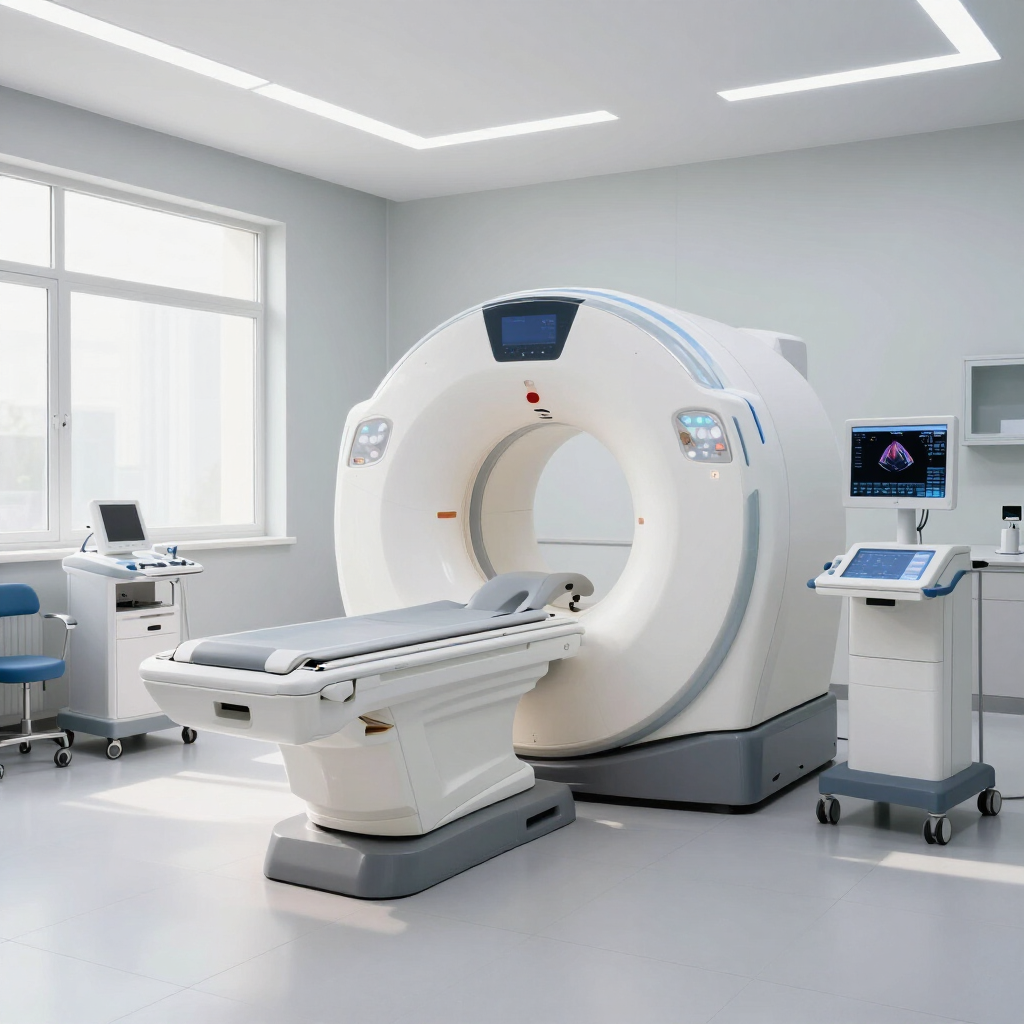
Ultrasound Scans Explained: What Happens During Your Scan
November 10, 2022
Ultrasound Scans:
Ultrasound scans, also called sonography, has helped facilitate significant improvements in the diagnosis of certain conditions. Throughout the years, ultrasound scanning applications have significantly grown. Ultrasound scan for pelvis, abdominal scan, and pregnancy are just some of the most common uses for this imaging procedure.
In part, the still-growing use of ultrasound is due to its proven effectiveness in supporting the accuracy of diagnosis across different medical fields. But. more importantly, its wide use and acceptance is also because it is non-invasive, delivers fast results, and the cost for ultrasound scan continues to be inexpensive.
Above all, ultrasound is perfectly safe, even for pregnant women such that there is no reason why a patient and the attending healthcare provider should withhold the procedure. Ultrasound is not associated with any serious side effects unlike other imaging procedures, like x-rays, which exposes the patient to radiation.
How does ultrasound scan work?
Ultrasound imaging uses ultra-high frequency sound waves, that is, over 20 Khz, to bounce it against an area of the body under observation. If you will be undergoing this procedure, expect the physician or technician to use a transducer, which delivers the ultrasound, and apply gel over the area to be examined which serves to enhance the reception of soundwaves.
When the soundwaves are reflected back, these waves carry useful information which the machine then utilizes to create an image of various anatomical structures. Thus. the major advantage of this procedure is that it is able to produce an image of how the body looks inside without requiring the patient to be opened up for diagnosis.

Depending on the machine used, the image produced can either be a thin cross-section of the body or, a 3D image, which is currently the most popular ultrasound for pregnancy.
What is ultrasound scan used for?
Medical ultrasound can be categorised into either Therapeutic Ultrasound or Diagnostic Ultrasound. You may think of the first as the non-invasive alternative to surgery. On the other hand, an ultrasound scan will generally fall under the latter category.
When used for diagnostic purposes, an ultrasound scan is typically carried out to examine internal organs non-invasively. It must be stressed that ultrasound works poorly for producing scanned images of the bones or lungs.
In general, ultrasound scanning is used for numerous applications. So, what does an ultrasound scan show? Well, that would depend on what specific ultrasound scan is being undertaken. The most common ultrasound scan types are as follows:

- Pelvic Ultrasound: This procedure is typically done for women. It produces images of key reproductive organs, including the vagina, uterus, cervix, ovaries, and fallopian tubes.
- Abdominal Ultrasound: The scan produced from this procedure shows the liver, spleen, kidneys, pancreas, spleen, as well as major blood vessels in and around the area.
- Liver Ultrasound: An ultrasound scan liver can aid in the diagnosis of fatty liver, and also show structural damages to this organ.
- Rena or Kidney: Ultrasound. An ultrasound scan kidney aids in the assessment of the physical characteristics of the kidneys. It can show fluid build-up as well as the formation of kidney stones.
- Thyroid Ultrasound: The resulting scan shows nodules on the neck. Further, the image helps in the proper diagnosis of cysts and tumors in the neck area.
- Carotid & Abdominal Aorta Ultrasound: This procedure is typically recommended for men over 60 years who ever smoked to rule out abdominal aortic aneurysm.
- Vascular Ultrasound: Their procedure is used to examine blood flow throughout the body by taking images of veins and arteries. It is commonly accompanied by a Doppler ultrasound for a more accurate diagnosis.
- Transvaginal Ultrasound: This is a minimally invasive procedure where the transducer is inserted in the vagina to examine any the physical properties of the vagina, uterus, cervix, ovaries, and fallopian tubes.
- Transrectal Ultrasound: Usually performed on adult men, this procedure involves inserting the transducer into the rectum to examine the rectum and prostate.
- Obstetric Ultrasound: In obstetric imaging, the primary objective is to evaluate the growth of the fetus. An OB-GYN also uses the information from a pregnancy scan to assess the overall health and safety of pregnancy. The overarching objective is the safety of pregnancy and delivery for both the mother and child. Among other things, an OB ultrasound imaging can help:
- Estimate the age of pregnancy and date of delivery
- Determine the position of the fetus
- Show fetal developmental abnormalities
- Ascertain abnormal pregnancies, such as placenta previa
- Show multiple pregnancies
Can ultrasound scan detect cancer?
An ultrasound can help in the detection of tumors, cysts, and abscesses. It can also show any barriers, abnormal collection of fluids such as in the kidneys, and signs of inflammation which, in turn, may signify underlying causes such as infection or cancer.
However, where a physician suspects the presence of cancer, the patient will typically be made to undergo a series of tests to improve the accuracy of diagnosis. In fact, an ultrasound promotes precision when a physician needs to take a tissue sample for biopsies.
However, it must be stressed that an ultrasound alone cannot provide sufficient information to properly screen for cancer.
However, it must be stressed that an ultrasound alone cannot provide sufficient information to properly screen for cancer.
Where To Get Private Ultrasound Scan?
Before you Google, “ultrasound scan near me,” take the time to check out the imaging services available at MRIPlus. All of the ultrasound procedures discussed here can be performed in the clinic as outpatient procedures.
For early pregnancy scans private and other ultrasound procedures, call MRIPlus for appointments and further inquiry.
For early pregnancy scans private and other ultrasound procedures, call MRIPlus for appointments and further inquiry.
References:
Public Health England. Guidance.Ultrasound: what is is, how it works and impact of exposure. Link:
https://www.gov.uk/government/publications/ultrasound-what-it-is-how-it-works-and-the-impact-of-exposure/ultrasound-what-it-is-how-it-works-and-impact-of-exposure Access Date: 29 Jul 2020
https://www.gov.uk/government/publications/ultrasound-what-it-is-how-it-works-and-the-impact-of-exposure/ultrasound-what-it-is-how-it-works-and-impact-of-exposure Access Date: 29 Jul 2020
National Institute of Biomedical Imaging and Bioengineering. Ultrasound. Link:
https://www.nibib.nih.gov/science-education/science-topics/ultrasound Access Date: 29 Jul 2020
https://www.nibib.nih.gov/science-education/science-topics/ultrasound Access Date: 29 Jul 2020
Ter Haar, Gail. Ultrasonic imaging: safety considerations. Interface Focus. Interface Focus. 2011 Aug 6; 1(4): 686–697. The Royal Society Publishing. Published online 2011 May 25. doi: 10.1098/rsfs.2011.0029. Link:
https://www.ncbi.nlm.nih.gov/pmc/articles/PMC3262273/ Access Date: 29 Jul 2020
https://www.ncbi.nlm.nih.gov/pmc/articles/PMC3262273/ Access Date: 29 Jul 2020
By Rahul Panchal
•
July 28, 2026
Discover the critical red flags for headaches in an MRI. Learn about warning signs like thunderclap onset and neurological symptoms that require urgent brain imaging.
By Rahul Panchal
•
July 27, 2026
Learn how to diagnose pancreatic cancer, recognize early symptoms, and understand the role of MRCP MRI scans in early detection and screening for high-risk patients.
By Rahul Panchal
•
July 24, 2026
Learn more about the thoracic spine anatomy, common conditions, and what to expect from a thoracic spine MRI. Middle back pain: What our experts say about signs and treatment.

By Rahul Panchal
•
July 23, 2026
Discover why removing metal is essential for MRI safety. Learn how MRI machines work, the risks of metallic objects, and how to prepare for your private scan.


