

If you are searching for an MRI scan near me, you probably want two things: speed and clarity. MRI Plus helps patients book private diagnostic imaging with straightforward self-pay options, referral guidance, and digital results delivery. That matters when pain, injury, or unanswered symptoms are affecting daily life and you do not want to wait in uncertainty. A private MRI scan near me can be a practical next step when you need timely imaging and a report your clinician can act on. For a…
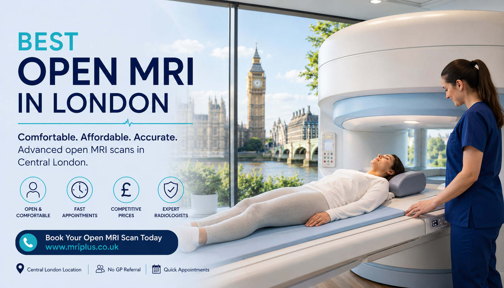
We offer both open MRI scanning across our London locations, with self-referral options so you don't need to wait on a GP appointment to get started.

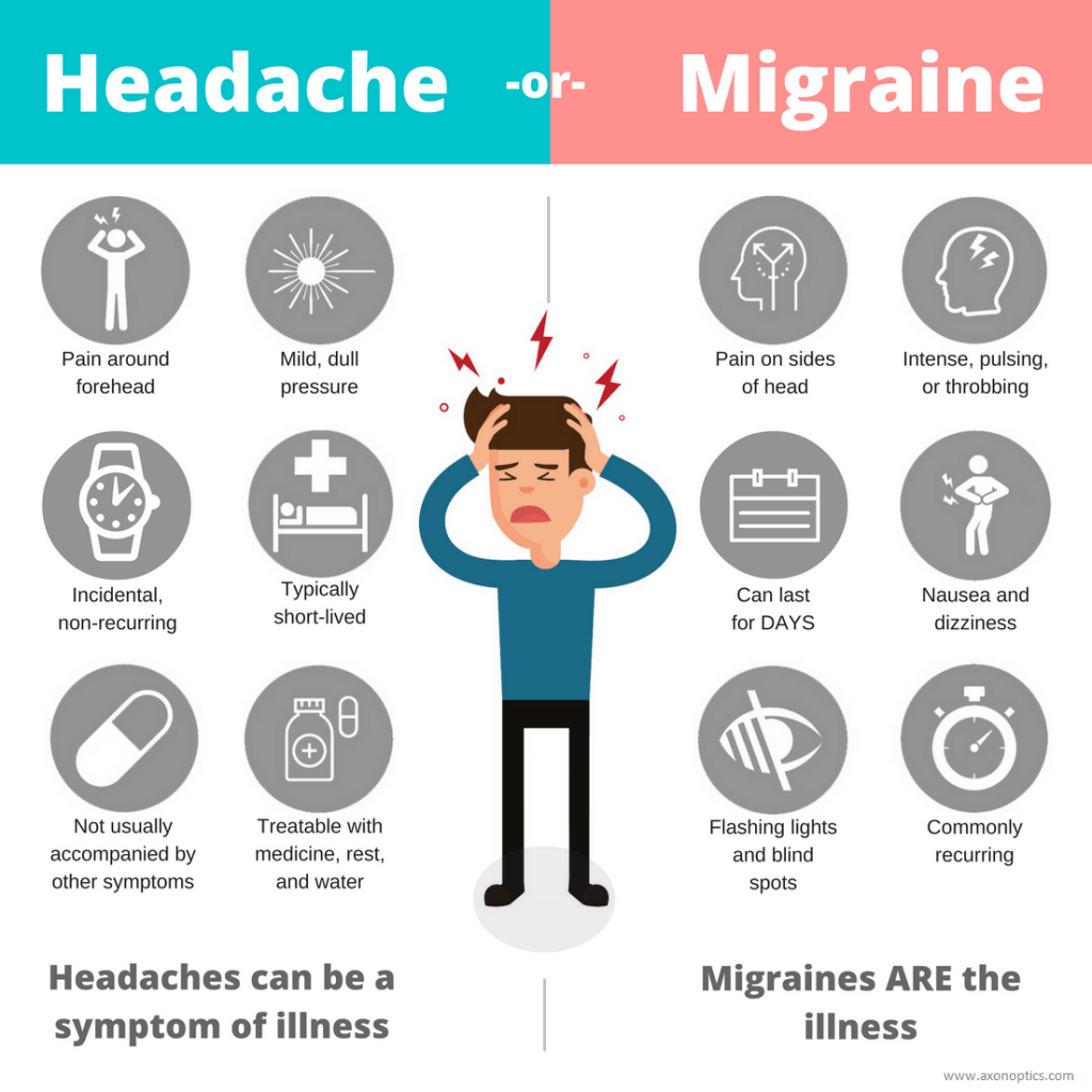
Discover the critical red flags for headaches in an MRI. Learn about warning signs like thunderclap onset and neurological symptoms that require urgent brain imaging.
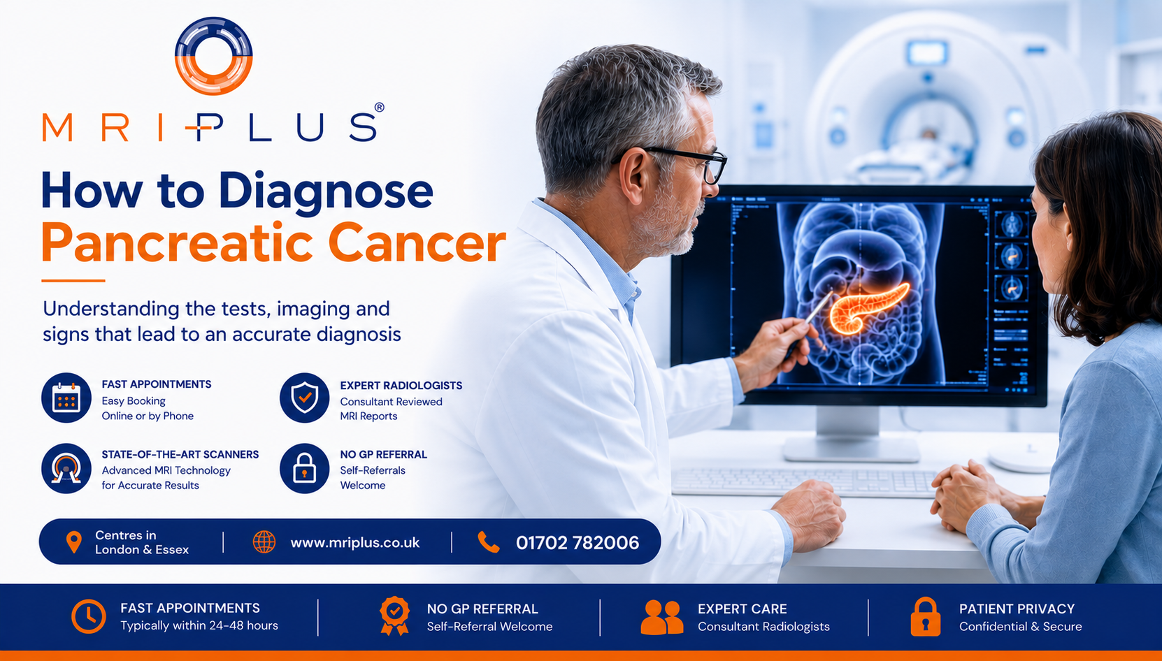
Learn how to diagnose pancreatic cancer, recognize early symptoms, and understand the role of MRCP MRI scans in early detection and screening for high-risk patients.


